Skip to main content
Dr Harsh Shah · Gastro & Cancer Surgeon · Apollo Hospital, Bhat, Gandhinagar and Gota OPD, Ahmedabad
Bleeding from the lower gut means blood passed from the back passage coming from the large intestine, or from the small intestine beyond the reach of an ordinary scope. Most of it stops on its own and almost none needs an operation — but it always needs an answer, the exact place the blood came from, because the worst outcome here is a bowel operation done on a guess. Dr Harsh Shah manages it at Shah’s Gastro, Cancer & Robotic Surgery Centre in the order the evidence now supports: resuscitate, decide whether the source is above or below, find the bleeding point, treat it through a scope or a catheter, and operate only on a located target.

Shah’s Gastro, Cancer & Robotic Surgery Centre
Dr Harsh Shah
MS, MCh (Surgical Gastroenterology), DrNB (Surgical Gastroenterology)
Registered with Gujarat Medical Council, Reg. No. G-38738
Gastro & Cancer Surgeon
· Apollo Hospital, Bhat, Gandhinagar · Gota OPD, Ahmedabad
Qualified twice over in surgical gastroenterology and trained in high-volume units, Dr Harsh Shah is the surgeon families are sent to when bleeding has not settled, when two scopes have found nothing, or when a bowel resection has been advised without a located source — work that is as much about preventing an unnecessary operation as performing a necessary one. Google rating 4.89 from 216 reviews at the Gota clinic, and 4.94 from 17 at the Apollo Hospital, Bhat listing.
Fresh or dark blood passed from the back passage usually comes from the colon — diverticular disease, fragile vessels in the bowel wall called angiodysplasia, a segment of colon with a reduced blood supply, colitis, or the raw area left after a polyp is removed. When the colon and the stomach are both clear, the source is most often the small intestine — now called small bowel bleeding rather than the older term obscure bleeding, a change of name that matters because a large share of these turn out to be lesions a standard scope could have reached.
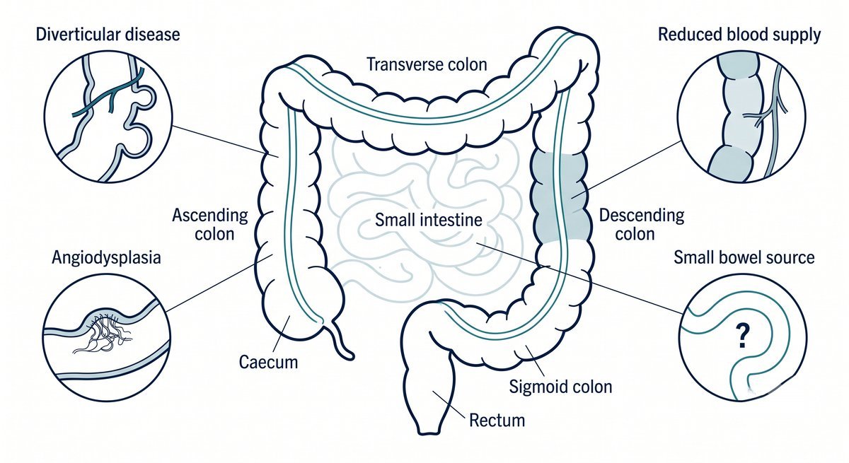
Two situations are handled elsewhere. Bleeding from varices in liver disease has its own pathway. And piles, fissure and fistula are not treated in this unit at all — referred to a colorectal service, with one strict condition attached.
An anorectal source is never accepted as the explanation for anaemia or significant blood loss until the colon above it has been examined. Piles are so common that a patient with a colonic lesion very often has them too, and blaming the visible thing is how a treatable lesion is missed for a year. In anyone past middle age, or with iron deficiency, weight loss or a change in bowel habit, the colon is evaluated fully whatever the back passage shows.
Fresh red blood from below, in a patient whose pulse is racing and whose blood pressure is falling, is a bleed from the stomach or duodenum until proven otherwise — a brisk upper gut haemorrhage travels through the intestine fast enough to arrive as fresh red blood. These patients need an upper endoscopy first. Sending them for bowel preparation instead wastes the hours in which the bleeding point could have been found and closed.
It is the one error here that turns a survivable event into a catastrophe, and it is why Dr Harsh Shah’s first question on any referral is not what the colonoscopy showed but what the pulse and blood pressure were doing when the blood appeared.
Which test comes first is decided by how stable the patient is, not by what is convenient to arrange. An unstable, actively bleeding patient goes to CT angiography, which shows the blood escaping and marks the spot for the radiologist to block the vessel. A stable patient whose bleeding has slowed goes to a properly prepared colonoscopy — not one done in the middle of the night.
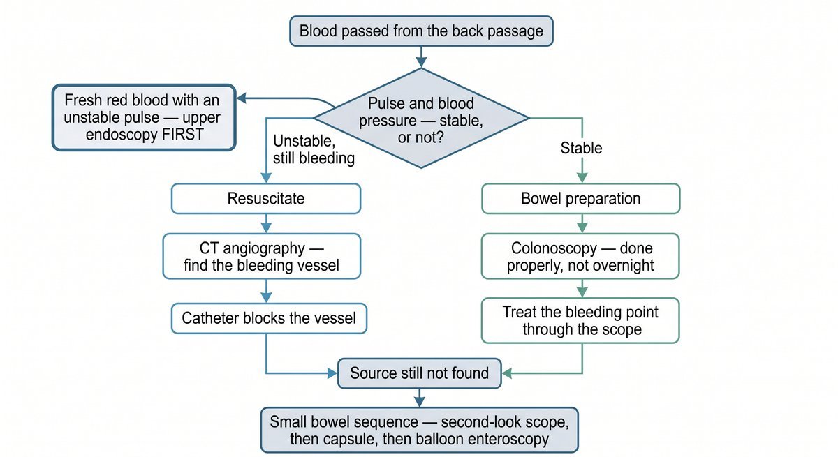
| The situation | What is done first |
|---|---|
| Unstable, still bleeding | Resuscitation, then CT angiography, then catheter angiography with blocking of the bleeding vessel |
| Fresh red bleeding with an unstable pulse | Upper endoscopy first — treated as a stomach or duodenal bleed until excluded |
| Stable, bleeding stopped or slow | Colonoscopy after proper bowel preparation, at a sensible hour |
| Colonoscopy and gastroscopy both clear | The small bowel pathway — second-look scope, capsule, balloon enteroscopy |
| Iron-deficiency anaemia, no visible bleeding | Full evaluation of colon and stomach before anything is attributed to piles |
Alongside the imaging, the first consultation records what decides management as much as the scan does: haemoglobin and its trend, clotting, kidney and liver function, and the list of aspirin, clopidogrel, warfarin and newer blood thinners with the reason each was started. That last item is not administrative detail — it changes what is safe to do next.
Until recently the teaching was that a high-risk patient with lower gut bleeding should have a colonoscopy within a day. The American College of Gastroenterology reversed that in its updated guideline: doing it urgently has not been shown to reduce rebleeding or improve any outcome that matters, and in a stable patient a colonoscopy within the next fortnight is as effective as one done overnight. A well-prepared colon examined in daylight is safer and finds more.
Families find this counter-intuitive, and they are entitled to an explanation rather than an instruction. An overnight colonoscopy after a rushed preparation looks through a colon still full of blood: the bleeding point is missed and the procedure usually has to be repeated. Waiting is not delay — it is the difference between a diagnostic procedure and a wasted one.
The exception is the unstable patient, and there the answer is not endoscopy at all. Someone bleeding actively with a failing circulation goes to CT angiography and then to the interventional radiologist, who threads a catheter to the vessel and blocks it. A scope in that situation is the wrong instrument at the wrong moment.
Most lower gut bleeding is controlled without surgery. At colonoscopy a bleeding point can be clipped, injected or banded, and fragile vessels in the bowel wall sealed with argon plasma. Where bleeding is brisk and has been pinpointed on CT, the interventional radiologist blocks the feeding artery from inside. Each treats the lesion where it lies and leaves the bowel intact.
Diverticular bleeding is the commonest cause and the most reassuring one: usually without pain, often alarming in volume, and stopping by itself in the large majority of episodes. Where a bleeding point is seen it is treated through the scope; where bleeding continues and has been localised, the vessel is blocked. Only repeated, localised, transfusion-dependent bleeding from one identified segment argues for removing that segment.
Angiodysplasia is often managed better with patience than with instruments. These fragile vessels recur, so in an older or frail patient long-term iron replacement frequently serves better than repeated procedures. They are also associated with a tight aortic valve and with kidney disease, so the heart and kidney function are reviewed rather than treated as coincidences.
A colon segment with a poor blood supply usually settles with supportive care; surgery is reserved for bowel that has lost its blood supply, for peritonitis, or for a narrowing left behind. Bleeding after polyp removal is treated at colonoscopy. Colitis is treated as the underlying condition rather than as a bleeding problem.
Blood thinners are handled with the doctor who prescribed them, never unilaterally, and anticoagulation restarts once bleeding is secure: the risk of a clot during a long interruption is real and consistently under-weighted next to the visible fear of more bleeding.
When the colon and the stomach are both clear, the sequence is fixed and worth following in order. A second look with an ordinary scope comes first, because a substantial share of small bowel bleeds are lesions within reach that were missed. Then a video capsule — a swallowed camera — but only once a narrowing has been excluded, since a capsule stuck in a stricture has to be retrieved; CT enterography comes first where obstruction or a mass is suspected. Balloon enteroscopy then reaches the lesion to treat it and mark it, so a surgeon can find it later.
Two additions belong in specific situations. During active bleeding, CT angiography or a red-cell scan localises the segment while it is visible. In a young adult with unexplained bleeding, a Meckel scan looks for a pouch left over from development — missed for years because nobody thinks of it in an adult.
The last resort is enteroscopy performed during the operation itself, surgeon and endoscopist working together. It exists so that a patient already on the table still gets a located source rather than a guessed resection.
Surgery is the last step, and localisation comes before it. A colonic bleed pinned to one identified segment, that keeps returning and keeps needing transfusion, is treated by removing that segment; a small bowel lesion marked at enteroscopy is removed with a short length of intestine. Both are operations aimed at a target. What is never done is a blind removal of part of the colon in a patient whose bleeding point has not been located.
The reason is the most important sentence on this page. If the source was never found, the chance that it lies in the colon left behind is unacceptably high — the patient bleeds again, having been put through a major operation, and is weaker for the second one. When rebleeding follows a segmental resection, the usual explanation is not bad luck but that the wrong segment was taken.
One situation does go ahead without a located source, as a deliberate decision rather than a default. A patient bleeding torrentially, in whom every attempt at localisation has failed, is treated by removing the whole large intestine and bringing out an ileostomy. It is a big operation in an unwell patient, it carries a real risk to life, and it is chosen consciously with the family told exactly why — which is not the same thing as guessing at a segment.
The approach — keyhole or open — follows the patient’s condition rather than preference: a planned, localised resection in a stable patient is usually laparoscopic, while an unstable patient bleeding heavily is operated on in the way that is fastest and safest, generally open. Dr Harsh Shah counsels every complication by name beforehand: leakage from the join, a stoma that may be temporary or permanent, infection inside the abdomen, slow return of bowel function, adhesions, a hernia at the wound, and bleeding again if the source was not correctly identified. The figures for your own case are given at the consent discussion in person, not quoted on a web page.
Discharge is not decided by the calendar. It needs a stable circulation, a haemoglobin that is holding, no ongoing bleeding — and either a diagnosis or a written plan to reach one. Sending a patient home with unexplained gut bleeding and no arranged next step is the commonest failure in this condition, and it is preventable.
Four things are checked before anyone leaves: the diagnosis, or the named investigation booked with a date; the blood-thinner plan, with the doctor who owns it; iron replacement, which is forgotten routinely; and written red-flag instructions — bleeding again, dizziness, fainting or a racing pulse means coming back rather than waiting for the review appointment.
Patients who have had an operation follow the standard enhanced-recovery route — eating early, out of bed on the first day, stoma teaching begun in hospital. Review follows at about a month to six weeks with haemoglobin and iron studies, to confirm the anaemia is correcting and that every outstanding investigation was completed. Investigations arranged at discharge and never chased are the other half of the same failure.
An admission for gut bleeding is usually an emergency, so cashless pre-authorisation is opened by the office at Apollo Hospital, Bhat as soon as the admission is made; CGHS and corporate panels go through the same office. An itemised written estimate follows once the plan is clear. What moves it is transfusion, whether CT angiography and embolisation are used, how many endoscopic procedures are needed, whether an operation follows, room category and length of stay — so a single figure quoted over the phone is a guess.
★★★★★
“I am writing to express my sincere gratitude for the exceptional care provided by Dr. Harsh Shah and his team. Dr. Harsh Shah is an outstanding expert in intestinal surgery, executing complex procedures with remarkable calm and precision. Dealing with gastrointestinal diseases is incredibly stressful for both patients and their families. In my experience, what sets Dr. Harsh Shah apart is his incredible patience; he takes the time to listen to every question and provides clear, reassuring answers to every concern.”
★★★★★
“I done my stoma closer operation with harsh sir such a nice and kind person never doing miss guide always give right advice polite nature.”
★★★★★
“I had my appendix surgery done by Dr. Harsh Shah, and I am very satisfied with the care I received. Dr. Harsh Shah explained the procedure clearly, performed the surgery smoothly, and ensured I was comfortable throughout my treatment. The recovery has been excellent, and the entire experience was reassuring. I highly recommend Dr. Harsh Shah for his professionalism, expertise, and compassionate care.”
— Dipen Shah, Google review, Apollo Hospital, Bhat listing
— Jigar Jadav, Google review, Gota clinic listing
— Sakina Magar, Google review, Apollo Hospital, Bhat listing
Yes. Stopping on its own is the usual behaviour of this bleeding, not the all-clear. The episode still needs a cause: the same lesion can bleed again, and a small number come from something that must not be left undiagnosed. What changes once the bleeding stops is the urgency, not the need.
Because doing it urgently does not improve the outcome and makes the test worse. The updated American College of Gastroenterology guidance reversed the older advice: in a stable patient a properly prepared colonoscopy is as effective as one done overnight, and it is safer and far more likely to find the bleeding point. An unstable patient is different — there the next step is CT angiography, not a scope.
Only if the colon above has been examined, and it is never enough to explain iron-deficiency anaemia. Piles are so common that a patient with a colonic lesion usually has them too. Piles, fissure and fistula are not treated in this unit and are referred to a colorectal service — but anyone with anaemia, significant blood loss, weight loss, a change in bowel habit, or past middle age has the colon evaluated first.
You move to the small bowel pathway, in order: a second look with an ordinary scope, because a large share of these lesions are within reach and were missed; then a video capsule, once a narrowing has been excluded; then balloon enteroscopy to reach the lesion, treat it and mark it. Skipping steps is what leaves patients circling for months.
Most patients need neither. Surgery is done only on a located source, usually a short segment. A stoma becomes likely only in the emergency where the whole large intestine must be removed in someone bleeding heavily with no source found. If an operation is proposed without a located bleeding point, that is the moment to ask for a second opinion.
Shah’s Gastro, Cancer & Robotic Surgery Centre
Gota, Ahmedabad, Gujarat
Google rating 4.89 from 216 reviews
Dr Harsh Shah consults at two addresses — Gota, Ahmedabad, and Apollo Hospital, Bhat.
Passing blood, or told your haemoglobin is low with no source found? Send the colonoscopy report, the CT images themselves and the list of blood thinners being taken — that combination usually settles the next step. Call +91-63555-64601 · WhatsApp Dr Harsh Shah's office
Swasth Parivar: every week, practical advice on recovery, diet and warning signs. No advertising. Stop any time.
Your number is used for these updates and for the clinic to reach you. It is never sold or passed on. To stop, reply STOP on WhatsApp.