WhatsApp Skip to main content

Dr. Harsh J Shah

Ulcerative Colitis: Medicines That Control It, the Day-3 Rule in a Severe Flare, and Surgery That Removes It

Dr. Jivraj Mehta Hospital · Vasna, Ahmedabad

Tapping Book on WhatsApp will likely open the WhatsApp app on your phone.

Ulcerative colitis treatment in Ahmedabad · Dr. Jivraj Mehta Hospital, Vasna, Ahmedabad

Most people with ulcerative colitis are treated with medicines and never need an operation. Surgery to remove the colon is a definitive treatment, not a failure.

Ulcerative colitis (UC) is an inflammation of the lining of the large bowel. It starts in the rectum. It spreads upwards without gaps. It causes bloody diarrhoea with mucus and an urgent need to pass stool. Mesalamine-type medicines control most of it. They are taken by mouth and as enemas or suppositories. Steroids treat flares. Newer drugs that calm the immune system are added when these are not enough. A severe flare needs hospital care on a strict day-by-day timetable. Once the colon is removed, the colitis in it is gone.

Ulcerative colitis, Crohn's disease, or an infection?

Bloody diarrhoea has more than one cause, and each is treated differently. UC must be told apart from Crohn’s disease, bowel infections and, in this region, bowel tuberculosis.

TB of the bowel can look like colitis. It becomes dangerous if drugs that suppress the immune system are started without checking for it.
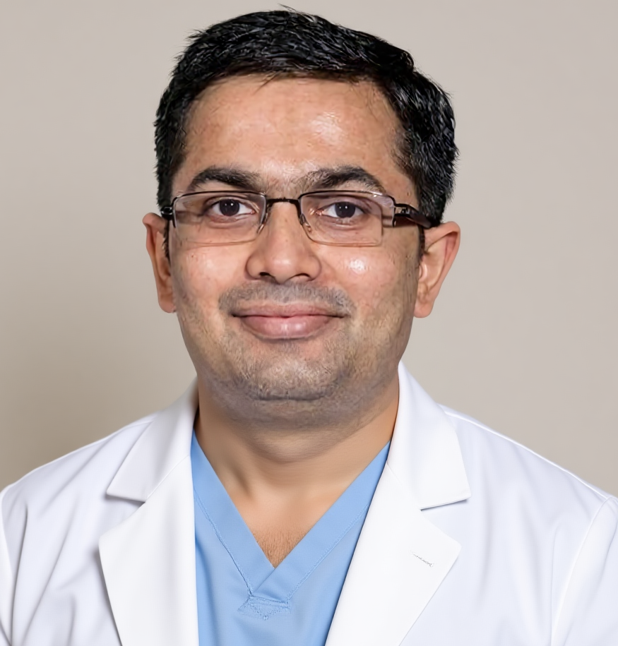
Dr Harsh Shah, laparoscopic colon surgeon, Ahmedabad
Dr Harsh Shah — MS, MCh (Surgical Gastroenterology), DrNB (Surgical Gastroenterology).

Dr. Jivraj Mehta Hospital. Dr Harsh Shah trained in super-specialty surgical gastroenterology programmes. He is qualified twice over in the specialty. He has 18 years of experience in bowel surgery. One practice covers the whole path. That includes colonoscopy review, and medical treatment planned with gastroenterology. It includes joint care of severe flares from the first day of admission. It also includes emergency colectomy, planned pouch surgery and permanent ileostomy, stoma care with a stoma nurse, and long-term surveillance.

Dr Harsh Shah
MS, MCh (Surgical Gastroenterology), DrNB (Surgical Gastroenterology)
Registered with Gujarat Medical Council, Reg. No. G-38738 — Gastro, Cancer & Laparoscopic Surgeon
Dr. Jivraj Mehta Hospital, Vasna, Ahmedabad

Last updated: 10 October 2026.

Google rating 4.94 from 18 reviews at Dr. Jivraj Mehta Hospital, Vasna, Ahmedabad.

The usual story is bloody diarrhoea with mucus. There is an urgent need to reach the toilet. There is a feeling that the bowel has not emptied. Many wake at night to pass stool. A severe attack brings fever, a fast pulse, weight loss and anaemia. Some patients also have aching or swollen joints. Others have painful red lumps on the shins, skin ulcers, or red and painful eyes. Some have abnormal liver tests. These matter. They can point to a bile-duct condition called primary sclerosing cholangitis (PSC). PSC changes how closely the colon is watched in later years.

What else can cause bloody diarrhoea, and how it is told apart.
ConditionHow it is told apart
Ulcerative colitisInflammation with no gaps, starting in the rectum and spreading upwards. It is seen at colonoscopy and confirmed on biopsies.
Crohn’s disease of the colonPatchy inflammation, often at the end of the small bowel. Biopsies from the rectum and the last part of the small bowel tell the two apart.
Bowel infection, including C. difficileStool cultures and a C. difficile toxin test. These are checked at diagnosis and again at every flare.
Cytomegalovirus (CMV) in a flare not settling on steroidsBiopsies tested for CMV.
Tuberculosis of the intestineBiopsies. TB screening before any drug that suppresses the immune system.

Ulcerative colitis is not a cancer — but the colon is watched

UC is a benign, inflammatory disease. Over many years, long-standing and extensive colitis raises the risk of pre-cancerous change (dysplasia) in the colon lining.

That is why colonoscopy is repeated on a schedule, years after diagnosis. This is not because the patient has cancer. It is so that any change is found early. The risk rises with how long the colitis has been present. It rises with how much of the colon is involved and how severe the inflammation has been. It is higher when PSC is also present. Keeping the inflammation controlled on mesalamine-type medicines is itself protective. The surveillance schedule is set out further down, under follow-up.

The tests, in the order they are done

Colonoscopy with biopsies makes the diagnosis and shows how far the colitis extends. Stool tests rule out infection. Blood tests measure inflammation and anaemia.

Biopsies are taken from several segments, including the rectum and the end of the small bowel. Before any drug that suppresses the immune system, TB and certain viral infections must be checked for. In Indian practice this is not optional.

Suspected ulcerative colitis — the tests and what each decides.
TestWhat it decides
Colonoscopy with segmental biopsies, including rectum and end of the small bowelThe diagnosis, how far the colitis extends, and whether it is UC or Crohn’s disease.
Stool cultures, C. difficile toxin, ova and parasitesWhether an infection is causing or worsening the diarrhoea.
Blood count, CRP, ESR, albumin, iron studies, liver testsHow active the inflammation is. Anaemia and nutrition. Whether the bile ducts need checking.
Faecal calprotectinA stool marker of bowel inflammation. It is used again later to monitor treatment.
Chest X-ray and IGRA or Mantoux testTB, which must be checked before drugs that suppress the immune system.
Hepatitis B and C, HIV, chickenpox status, vaccination reviewInfections that can flare when the immune system is suppressed.

In a severe flare the order changes. A plain X-ray of the abdomen looks for a dangerously widened colon. A short, gentle look at the lower bowel confirms how severe it is. This is done without bowel preparation and is called flexible sigmoidoscopy. It also takes biopsies for CMV. A full colonoscopy is not done during a severe attack, because the inflamed bowel can perforate (tear through). A CT scan is added when a tear or toxic widening is suspected.

How far it extends, and how severe the flare is

Two things guide treatment: how much of the colon is inflamed, and how severe the current flare is.

Inflammation only in the rectum is treated very differently from colitis of the whole colon. A severe flare is a medical emergency that needs admission.

Extent of ulcerative colitis (Montreal classification).
ExtentWhat it means
ProctitisOnly the rectum is inflamed.
Left-sided colitisInflammation from the rectum up to the bend of the colon under the spleen.
Extensive colitis (pancolitis)Inflammation beyond that bend, often the whole colon.

A flare is called acute severe ulcerative colitis when two things are present. There are six or more bloody stools a day. There is also at least one sign that the whole body is affected: fever, a fast pulse, a low haemoglobin, or raised inflammation markers in the blood. These are the Truelove and Witts criteria. Every patient admitted with a severe flare is checked against them on the day of admission.

The treatment ladder for ulcerative colitis

Treatment is matched to extent and severity. Mesalamine-type medicines (5-ASA) work in UC, unlike in Crohn’s disease, and are continued long term.

Steroids treat flares but are never a long-term answer. Immune-modifying tablets and newer advanced drugs are used when steroids cannot be stopped. Surgery is the last rung for some. For a few, it is the first choice.

From enemas to an operation — who is treated how.
SituationUsual treatment
Proctitis or left-sided colitis, mild to moderateMesalamine suppositories or enemas first, with mesalamine tablets. Local treatment works better in the lower bowel than tablets alone.
Extensive colitis, mild to moderateMesalamine by mouth and by enema. A course of steroid tablets if this is not enough.
Steroids cannot be stopped, or do not workThiopurine tablets, or an advanced therapy.
Acute severe flareHospital admission and steroids into the vein. Then a rescue drug or surgery, on a fixed timetable.
Disease that keeps returning, poor quality of life, dysplasiaA planned operation.

The advanced therapies include anti-TNF injections or infusions (infliximab, adalimumab, golimumab). They also include vedolizumab, ustekinumab, the IL-23 blockers, JAK-inhibitor tablets and S1P-modulator tablets. The choice depends on how severe the disease is and how fast a response is needed. Other illnesses, cost and availability matter too. The aim is not just feeling better. It is normal blood and stool markers, and in the end a healed lining at colonoscopy. That is what keeps patients out of hospital.

When surgery is not needed

For most people with ulcerative colitis, no operation is ever needed. Mild and moderate disease is controlled with medicines, monitored with stool and blood markers.

These are mesalamine, local treatment for the rectum and short steroid courses for flares. Where needed, immune-modifying or advanced drugs are added. A common reason for a flare is simply that mesalamine was stopped once the bleeding settled. It is a maintenance medicine. It also lowers the long-term risk of bowel cancer. So it is continued even when the patient feels well. Enemas and suppositories are under-used because they are a nuisance. Yet for disease in the lower bowel they work better than tablets alone. The cost of advanced drugs is discussed early and openly. Where they are not affordable, surgery is a fair, definitive choice, not a last resort. That is said plainly.

A severe flare: the hospital timetable

In a severe flare, the greatest danger is an operation done too late in a patient who is getting worse. So the surgeon is involved from day one.

The danger is not an operation done too early. Two dates are fixed in advance. Day 3 is when the response to steroids is judged. Day 7 is when the response to the rescue drug is judged.

  1. Day 1. Admission, with the colorectal surgeon told and sharing care from the start. Steroids into the vein. Stool tests for infection, including C. difficile. Blood-thinning injections to prevent clots. These are given even though there is rectal bleeding, because severe colitis carries a high risk of clots. Opioid painkillers and anti-diarrhoea medicines are stopped. They can trigger dangerous widening of the colon. A limited sigmoidoscopy takes biopsies for CMV. The stoma nurse sees the patient and marks a possible stoma site early. That way it is never done in a rush.
  2. Every day. A stool chart, examination, blood tests, and an abdominal X-ray if the abdomen swells.
  3. Day 3. The response is measured against the Oxford criteria: the number of stools and the CRP level. If the patient has not responded, the next step is a rescue drug or an operation. The rescue drug is infliximab or ciclosporin, which work about equally well. A few more days of steroids is not the answer.
  4. Day 5 to 7. If there is no clear gain on the rescue drug by day 7, the colon is removed. A second rescue drug is not tried after the first has failed.
  5. At any point. Toxic widening of the colon, a tear (perforation), heavy bleeding or a patient who is getting worse means an operation the same day.

What patients say

★★★★★

★★★★★

“He is simple and straight conversation person. Explains the procedure and process to prepare the patient mentally for the surgery which makes patients comfortable during the process. He is also very patient enough to hear all problems of his patients which makes him an approachable doctor. — palak thareja, Google review”

★★★★★

“Dr. Harsh Shah is an excellent doctor who handled our case with great diligence and care. He listens attentively and addresses every concern with utmost patience. He's very humble and answers all of your questions in detail which is very assuring. I would highly recommend Dr. Harsh Shah for consultation and treatment. — Srinath Vasudevan, Google review”

The reviews below come from Dr Harsh Shah’s two Google listings, and are quoted exactly as written.

None names ulcerative colitis. They describe his care of bowel and abdominal conditions in general.

Why waiting longer is the danger

Going on with steroids, or adding one drug after another, in a patient who is not responding raises the risks of the operation that is eventually needed.

A patient who reaches surgery after weeks of high-dose steroids, weight loss and a widening colon is weaker. That patient is more infected and slower to heal. Many patients and families resist surgery. They believe it means the doctors have failed, or that it will not help. In UC the reverse is true. Removing the colon removes the diseased organ, and it usually makes the patient well. This is a real difference from Crohn’s disease, where an operation does not end the disease. Saying this clearly at the start of a severe admission is part of the treatment. It should not wait until day 7.

The emergency operation: removing the colon, keeping the rectum

In an emergency the operation is always the same: subtotal colectomy with an end ileostomy. The colon is removed and the rectum is left in place.

The end of the small bowel is brought out as a stoma. The rectum is closed off. No join and no pouch are made in a patient who is acutely ill. Rebuilding is decided later, once the patient is well.

The emergency operation — what is done and why.
StepReason
The colon is removed.It removes the diseased bowel and the source of infection and bleeding.
An end ileostomy is made.A join in a patient loaded with steroids, infected and under-nourished is unsafe.
The rectum is kept, closed or brought out as a small opening.It keeps every future option open, including a pouch.
A tube is placed in the rectum through the back passage.It takes pressure off the closed rectum and protects against it bursting.
Keyhole surgery in a stable patient.Smaller wounds, with the same operation inside.

The stoma made in an emergency is not always permanent. Whether the bowel is later joined again is decided when the patient is well and off steroids. The final report on the removed colon must also be known. During the operation, the swollen and fragile colon is handled with great care. A tear during removal would spill bowel content into the abdomen.

Planned operations: a pouch, or a permanent ileostomy

A planned operation has two main options, and both are good ones. One is a pouch made from small bowel. The other is a permanent ileostomy.

A planned operation is considered for colitis that keeps returning despite treatment. It is also considered for steroid dependence, a quality of life that cannot be borne, and dysplasia found at surveillance. It is also used to rebuild the bowel after an emergency colectomy. The pouch is joined to the anus. The other option removes the colon and rectum and leaves a permanent ileostomy.

Planned operations for ulcerative colitis.
OperationWho it suits
Restorative proctocolectomy with ileal pouch–anal anastomosis (IPAA)Patients with good control of the back passage who want to avoid a permanent stoma. It is usually done in stages, with a temporary ileostomy that is closed later.
Panproctocolectomy with end ileostomyWeak sphincter muscles, older patients, or patient choice. It is one reliable operation, and not a lesser choice.
Ileorectal anastomosisOccasionally, when the rectum is spared and there is no dysplasia. The rectum then needs regular checks.

Before a pouch, three things are confirmed. First, the disease really is UC and not Crohn’s disease, because a pouch in Crohn’s disease fails far more often. Second, the patient understands life with a pouch. That means several bowel movements a day and at night, and bouts of pouch inflammation. Third, for a woman who may want children, the effect of pelvic surgery on fertility has been discussed before the operation is booked. One choice is to delay the pouch until the family is complete.

Preparing for a planned operation

Preparation lowers the risks. Steroids are cut down as far as possible, nutrition and anaemia are corrected, and the stoma nurse marks the stoma site in every case.

The patient is counselled with written and visual information. Ideally, this comes after talking to someone who already lives with a stoma or a pouch.

  1. Steroid dose brought down as far as safely possible. A written plan covers steroids around the operation.
  2. Nutrition assessed and supplemented. Anaemia corrected.
  3. Stoma site marked by the stoma nurse, even when a stoma is not planned to be permanent.
  4. Blood-thinning injections to prevent clots, continued for four weeks after surgery.
  5. Antibiotics at the start of the operation. Keyhole surgery where suitable.

After the operation

Recovery follows an enhanced-recovery plan: sitting out and drinking on the day of surgery, then eating and walking from the first day.

Stoma care is taught from day one. Going home depends on the patient or a family member managing the stoma without help. That is usually the step that takes the longest.

After colectomy for ulcerative colitis — the usual day-by-day path.
DayWhat happens
Day of surgerySitting out of bed and clear fluids. Pain relief with few opioids. A written steroid plan.
Next dayDiet as tolerated and walking four times. Catheter removed. Stoma teaching begins, and stoma output is measured.
Second dayFull diet, pain relief by mouth, output controlled.
Third to fifth dayGrowing independence with the stoma. Readiness for home is checked.
Fifth to eighth dayHome once the stoma can be managed without help.

Before discharge, the steroid taper is written down and explained. This includes what to do if the patient becomes unwell while coming off steroids. Stoma output is kept under control, with loperamide if needed. When output is high, oral rehydration solution is used rather than plain water. The follow-up visit is booked to discuss the report on the removed colon and the question of rebuilding. Call at once for fever, a fast pulse, pelvic or belly pain, or a very high stoma output with dizziness.

The risks, named in words

An emergency colectomy in a patient on high-dose steroids carries more risk than a planned operation. The main risks are explained before consent.

They are clots, infection, problems with the closed rectum, and dehydration from a high-output stoma. For a pouch, the honest expectations are frequent bowel movements and bouts of pouch inflammation. The figures that apply to each patient are given at the consent talk.

Emergency colectomy. Bleeding that needs a transfusion. Wound infection. Clots in the leg or lung, a risk that UC itself raises. Anaesthetic problems. A stoma, which is certain. The closed rectum breaking down, with infection in the pelvis. An abscess in the abdomen. Slow return of bowel function. A high-output stoma with dehydration. Wound breakdown, and a further operation. There is a risk to life. It is higher when the colon has perforated, has widened dangerously, or surgery has been delayed. Pouch surgery. Pouch inflammation (pouchitis), usually settled with antibiotics. Several bowel movements a day and at night, some urgency and occasional night-time leakage. A leak from the join, with pelvic infection. A channel between pouch and vagina in women. Bowel blockage. Narrowing of the join. Pouch failure needing a permanent ileostomy. Reduced fertility in women. Less often, sexual problems after pelvic surgery.

Follow-up and surveillance colonoscopy

For anyone with a colon still in place, surveillance colonoscopy for dysplasia begins eight years after diagnosis in extensive or left-sided colitis.

Follow-up depends on where the patient is on the ladder. On medicines, stool and blood markers are checked every few months. After surgery, the stoma, steroid taper, nutrition and the rebuilding question are reviewed.

Follow-up in ulcerative colitis.
SituationWhat is checked
On medicinesFaecal calprotectin every three to six months. Blood count, CRP, albumin and iron. Whether medicines are being taken.
After colectomyStoma, completion of the steroid taper, and nutrition. The rebuilding talk, once well and off steroids.
Living with a pouchBowel frequency, pouchitis bouts and quality of life. Pouch endoscopy as planned.
Dysplasia surveillanceHigh-definition colonoscopy with dye spray or targeted biopsies, from eight years after diagnosis. It is repeated every one to five years, according to risk.
Primary sclerosing cholangitis as wellColonoscopy every year from the time that diagnosis is made.
Any dysplasia foundJoint discussion on removal through the colonoscope, or colectomy.

Long-term care also covers bone health after steroid courses, and vaccinations. It covers cervical screening for women on drugs that suppress the immune system. It covers iron, vitamin B12 and mood. Living with a disease that keeps coming back is hard, and that is part of the talk too.

Your treatment, from first visit to follow-up

  1. Book your consultation. Bring every colonoscopy and biopsy report, and stool and blood reports. Bring a list of medicines with the dates they were started and stopped. A severe flare with fever and many bloody stools goes straight to the emergency department at Dr. Jivraj Mehta Hospital, Vasna, Ahmedabad.
  2. Confirm the diagnosis. Colonoscopy and biopsies if not done. Infection ruled out. TB screening before any drug that suppresses the immune system.
  3. Treat to target. Mesalamine, local treatment and, where needed, advanced therapy, with gastroenterology. Treatment is adjusted by symptoms, markers and colonoscopy.
  4. Act on time in a severe flare. Joint care from day 1. Decisions on day 3 and day 7.
  5. Plan surgery when it is the better choice. Emergency colectomy, or a planned pouch or permanent ileostomy after counselling.
  6. Stay under surveillance. Colonoscopy on schedule for anyone with a colon in place.

Cost, insurance and admission

The cost of ulcerative colitis care varies more than for most conditions. Mesalamine and steroids are cheap, while advanced therapies are costly and continue for long periods.

An emergency admission and colectomy, and later rebuilding, are separate costs. These are discussed early. Cost is a real factor in choosing between long-term advanced therapy and a definitive operation. Dr. Jivraj Mehta Hospital, Vasna, Ahmedabad, works with the major insurers and cashless schemes. The office handles pre-authorisation directly, including emergency admissions. Bring colonoscopy and biopsy reports, stool and blood reports, and any CT or X-ray. Bring discharge papers from earlier admissions and a full list of medicines with doses. Bring the insurance card and identification.

Frequently asked questions

Will I have to take medicines for ulcerative colitis all my life?

Most people continue a maintenance medicine such as mesalamine for the long term, because stopping it is a common reason for a flare and it also lowers the long-term bowel-cancer risk. The dose and the need for other drugs are reviewed against symptoms, stool markers and colonoscopy.

Is a stoma after emergency surgery permanent?

Not necessarily. The emergency operation keeps the rectum so that a pouch can be considered later, once you are well, off steroids and the report on the removed colon is known.

Why was I given blood-thinning injections when I am passing blood?

A severe flare of ulcerative colitis makes clots in the legs and lungs more likely. Preventive injections are given despite rectal bleeding, because the clot risk is the greater danger.

Does removing the colon end ulcerative colitis?

Removing the colon removes the disease from the colon, which is a real difference from Crohn's disease. A pouch can later become inflamed (pouchitis), which is usually treated with antibiotics.

Can I have children after pouch surgery?

Pelvic pouch surgery reduces fertility in women. This is discussed before the operation is booked, and delaying the pouch until the family is complete is offered as an option.

Why do I need a colonoscopy every few years if I feel well?

Long-standing colitis raises the risk of pre-cancerous change in the colon lining. Surveillance colonoscopy from eight years after diagnosis finds such change early, when it can be dealt with.

Where Dr Harsh Shah sees and operates

Dr. Jivraj Mehta Hospital, Vasna, Ahmedabad
Dr. Jivraj Mehta Hospital, Dr Jivraj Mehta Marg, Vasna, Ahmedabad 380007.
Google: Dr Harsh Shah · 4.94 from 18 reviews
Directions to Dr. Jivraj Mehta Hospital, Vasna · WhatsApp +91-63555-64601

Patients come to Ahmedabad from across Gujarat — Gandhinagar, Mehsana, Banaskantha, Nadiad, Anand, Bhavnagar, Rajkot, Jamnagar and Kutch — and from southern Rajasthan and western Madhya Pradesh.

Diagnosed with ulcerative colitis, or told the colon may need to come out? Reports can be sent ahead of the visit. Send colonoscopy and biopsy reports, stool and blood reports, discharge papers and a list of medicines with dates. The consultation then starts with them already read. Treatment is decided at the visit, after examination. Six or more bloody stools a day with fever or a fast pulse need the nearest emergency department today. Call +91-63555-64601 · Bring the reports to the visit

Tapping Book on WhatsApp will likely open the WhatsApp app on your phone.

Get health updates from Dr Harsh Shah on WhatsApp

Swasth Parivar: every week, practical advice on recovery, diet and warning signs. No advertising. Stop any time.

Your number is used for these updates and for the clinic to reach you. It is never sold or passed on. To stop, reply STOP on WhatsApp.

Dr. Harsh J Shah
Dr Harsh Shah, Gastro, Cancer & Laparoscopic Surgeon, Ahmedabad
Privacy Overview

This website uses cookies so that we can provide you with the best user experience possible. Cookie information is stored in your browser and performs functions such as recognising you when you return to our website and helping our team to understand which sections of the website you find most interesting and useful.