Skip to main contentLast updated: 20-Nov-2026
Tapping Book on WhatsApp will likely open the WhatsApp app on your phone.

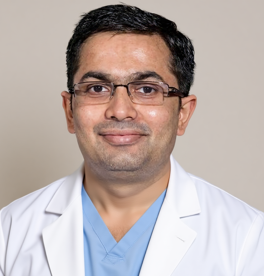
Dr Harsh Shah
MS, MCh (Surgical Gastroenterology), DrNB (Surgical Gastroenterology)
Registered with Gujarat Medical Council, Reg. No. G-38738
Liver & Gastro Surgeon
Dr. Jivraj Mehta Hospital, Vasna, Ahmedabad · 18 years of experience
In short
Most liver lumps are named by their pattern of blood flow. A good ultrasound comes first, then a multiphase CT or MRI that follows the contrast through the lump.
A special MRI with a liver-specific dye is kept for one hard question: telling a harmless scar-like lump from an adenoma, which may need an operation.
A needle test is the last step, not the first. It is used only when scans stay unclear and the answer would change the plan.
For the full picture of benign liver lumps, from the types to treatment choices, read our benign liver lesions treatment page. This post explains one thing only: which test tells you what the lump is.
Because every plan depends on the name of the lump. A harmless lump needs nothing, while a few types need a surgeon. The scan must answer two questions in order.
The first question is whether the lump is harmless or not. The second is which harmless lump it is. A haemangioma (a tangle of blood vessels) and a focal nodular hyperplasia, or FNH (a scar-like overgrowth), are simply left alone. An adenoma can bleed. A cyst with lumpy walls may be a growth. So the name decides everything that follows.
No single test answers both questions for every lump. Doctors therefore use a short, stepwise path. Each step is chosen to settle what the last step could not.
| Test | What it does well | Where it falls short |
|---|---|---|
| Ultrasound | Finds the lump, measures it and spots a classic haemangioma or simple cyst | Cannot name most solid lumps with certainty |
| Contrast ultrasound | Shows blood flow live, with no radiation and no strain on the kidneys | Looks at one lump at a time and depends on the operator |
| CT scan with contrast | Widely available and good at showing a haemangioma fill in | Uses radiation and is weaker at separating FNH from adenoma |
| MRI scan | Finest soft tissue detail, with fat and fluid checks | Costs more and may still leave FNH and adenoma close |
| MRI with liver-specific dye | The deciding test between FNH and adenoma | Costs more, takes longer and works less well in a sick liver |
It finds the lump, measures it and checks the rest of the liver. A bright, round, well-defined spot is often a haemangioma, and a clear fluid-filled sac is a simple cyst.
Ultrasound is cheap and needs no injection, so it is the right first test. It also shows whether the liver around the lump looks healthy, fatty or scarred. That background matters a great deal for the next step.
The limit is that ultrasound alone cannot name most solid lumps. A report that says “a lesion, advise further imaging” is not a bad sign. It only means the next test is needed.
When one lump needs a clear answer and a CT or MRI is not suitable. A tiny dose of bubble contrast is given in a vein and the lump is watched live.
The bubbles stay inside the blood vessels. They do not strain the kidneys and there is no radiation, so the test can be repeated. A haemangioma fills from its edge inwards and stays filled. A scar-like FNH fills quickly from the centre outwards. A harmful lump tends to empty of contrast early.
There is one trap. Some harmless lumps, adenomas especially, can also empty late. So a late wash-out on this test is not proof of anything. It is read together with the other scans.
They follow the contrast through the lump in stages. Each lump type has its own pattern, like a fingerprint. The order of the stages matters more than the size.
MRI also has a fat check. Fat inside a lump points towards one type of adenoma. This is why a good MRI often settles the question without anything more.
Because FNH and adenoma can look alike on ordinary pictures, yet one needs reassurance and the other may need surgery. A liver-specific dye separates them.
The dye is taken up by working liver cells and is held for about twenty minutes. An FNH has working liver cells, so it keeps the dye and stays bright on the late picture. An adenoma does not keep it and turns dark. That simple difference points to watching or to a planned operation.
This dye scan costs more and is not needed for every lump. It is kept for the one question that ordinary scans cannot answer.
By looking at its wall and what is inside. A simple cyst has a thin wall and clear fluid. Thick walls, partitions or lumps inside call for more care.
A cyst with a calcified wall, small cysts within it or a floating membrane may be a hydatid cyst. This is a parasite cyst, and it must never be punctured with a needle. A cyst with partitions and a lump on its wall may be a mucinous growth. That is removed, not drained. Our post on how scans tell a liver abscess from a hydatid cyst shows how doctors separate them.
Only when good scans stay unclear and the result would change the plan. A typical haemangioma, FNH or simple cyst is never sampled with a needle.
A needle into a blood-filled lump can bleed. A sample from an adenoma can also miss the one risky area. So doctors try to avoid it.
When it is done, the tissue is stained for a few markers. They sort an adenoma into its type, and each type has a different risk. A lump that stays doubtful, or sits in a risky spot, is sometimes removed to give a full answer.
Yes. In a scarred liver (cirrhosis), a new solid lump is read by stricter rules. The comfortable guess that it is probably harmless no longer applies.
FNH and adenoma mostly occur in livers that are otherwise healthy. If your liver is already scarred, a new lump needs a specialist read of the whole picture. Do not rely on a general report. A fatty liver mainly makes ultrasound harder to read. An MRI is then often the better choice.
Bring every scan report and, if possible, the actual scan discs. A surgeon can read the pictures himself, and that is better than reading only words.
No one scan suits every lump. Doctors begin with an ultrasound, then use a CT or MRI that follows contrast through the lump. A special MRI is added only for hard questions.
It means the first test found a lump but could not name it. This is common and is not a bad sign. The next scan is chosen to answer what the first one could not.
It is an ultrasound done after a small dose of bubble contrast is given in a vein. The bubbles show blood flow in the lump live. There is no radiation and the kidneys are not strained.
With a liver-specific dye. An FNH holds the dye and stays bright on the late picture. An adenoma does not hold it and turns dark. This guides the choice between watching and surgery.
No. A typical haemangioma, FNH or simple cyst is never sampled with a needle. A biopsy is used only when scans stay unclear and the result would change the plan.
Not before it is checked. A cyst with a thick or calcified wall may be a parasite cyst, and a needle can spread it. A scan and a blood test come first.
Yes. In a scarred liver, new lumps are read by stricter rules and need a specialist. Please do not rely on the general idea that most liver lumps are harmless.
For the full picture of benign liver lumps and their care, start at the benign liver lesions treatment page. To learn when such a lump is removed, read when a benign liver lump needs surgery. For questions before you travel, call +91-63555-64601.
Dr Harsh Shah — Dr. Jivraj Mehta Hospital, Vasna, Ahmedabad
Dr. Jivraj Mehta Hospital, Dr Jivraj Mehta Marg, Vasna, Ahmedabad 380007.
Consultations, review of reports and scans, operations and follow-up. OPD Monday to Saturday, 9 am to 5 pm.
Appointments: +91-63555-64601 · +91-63590-11009 · Hospital 079-2663 6363
Directions to the hospital · WhatsApp +91-63555-64601
A scan has shown a spot in your liver, and each report seems to say something different? Dr Harsh Shah, a liver and gastro surgeon in Ahmedabad, reads these scans with patients and decides which test is worth doing next. Bring every report and the scan discs. He will explain what the lump is likely to be and what, if anything, still needs checking. You can also call us or write on WhatsApp. Call For Appointment Book on WhatsApp Tapping Book on WhatsApp will likely open the WhatsApp app on your phone.
Tapping Book on WhatsApp will likely open the WhatsApp app on your phone.
Swasth Parivar: every week, practical advice on recovery, diet and warning signs. No advertising. Stop any time.
Your number is used for these updates and for the clinic to reach you. It is never sold or passed on. To stop, reply STOP on WhatsApp.