Skip to main content
Ahmedabad and Gandhinagar · Dr Harsh Shah, Liver Surgeon
Most lesions in the liver are found by accident, on a scan done for something else, and most need a confident diagnosis and then nothing at all — no operation, no repeat scans, no restriction on pregnancy or the contraceptive pill. The operations named above exist for the minority: an adenoma carrying a real risk, a cyst large enough to cause symptoms, and the one cystic lesion that must be removed whole rather than opened. The commonest harm here is not a missed diagnosis. It is a major liver operation done on a lesion that would never have troubled the patient.

Shah’s Gastro, Cancer & Robotic Surgery Centre
Dr Harsh Shah
MS, MCh (Surgical Gastroenterology), DrNB (Surgical Gastroenterology)
Registered with Gujarat Medical Council, Reg. No. G-38738
Liver Surgeon
· Apollo Hospital, Bhat, Gandhinagar · Gota OPD, Ahmedabad
Qualified twice over in surgical gastroenterology, and operating on the liver, pancreas and bile ducts as his daily work. A benign liver lesion is the one situation where the most valuable thing a liver surgeon can offer is an accurate reason not to operate. Dr Harsh Shah reads these scans himself alongside the radiologist, and tells patients plainly which of the six lesions they have and what follows from it. Google rating 4.89 from 216 reviews at the Gota listing, and 4.94 from 17 reviews at the Apollo, Bhat listing.
Six different things are meant when a report says a lesion, a mass or a cyst in the liver: haemangioma, focal nodular hyperplasia (FNH), hepatocellular adenoma, a simple hepatic cyst, polycystic liver disease, and a biliary mucinous cystic neoplasm. They behave completely differently. Two of them need nothing ever. One needs hormonal changes first and an operation only on defined grounds. One needs an operation only if it causes symptoms. One is managed jointly with a kidney specialist. And one has to be removed entirely, because opening it is the wrong operation.
What they have in common is only how they are found — an ultrasound for indigestion, a CT after an accident, a health check — and a report using the word tumour, which is the word that frightens people. Sorting out which of the six is present is nearly all of the work, and it is done with imaging rather than with a needle.
Before anything is said about the lesion itself, the liver around it has to be assessed. If the background liver is normal, the six conditions above are the realistic possibilities. If the background liver is cirrhotic or scarred, a new nodule in it is treated as a liver cancer until it is proved otherwise, and none of what follows on this page applies — that is a different condition, assessed on a different pathway, and it needs its own consultation.
Contrast-enhanced MRI, using a hepatobiliary contrast agent, is the single most useful test for a liver lesion and has the highest diagnostic performance of anything available. Its particular value is that it separates focal nodular hyperplasia from hepatocellular adenoma, which is the one distinction on which management genuinely turns. Multiphasic CT and contrast-enhanced ultrasound are reasonable alternatives, and when their typical features are present in combination they can identify FNH with near-complete confidence.
| Test | What it is actually being asked |
|---|---|
| Contrast MRI with hepatobiliary agent | Which of the six lesions is this — and specifically, is it FNH or an adenoma |
| Liver function tests, albumin, INR, blood count | Is the liver working normally, and is it safe to operate if an operation is needed |
| Hepatitis B and C serology, AFP where there is doubt | Is this actually a scarred liver, in which case the diagnosis changes entirely |
| The history — contraceptive pill, anabolic steroids, pregnancy, weight, alcohol | Is this an adenoma, and can it be made to shrink without an operation |
A lesion with typical haemangioma features is not biopsied. Biopsy is reserved for a lesion still genuinely indeterminate after good imaging, and then only after discussion at a joint meeting. Where the lesion is cystic, the task narrows to one question: is this an ordinary simple cyst, or the mucinous cystic neoplasm described below.
A haemangioma is the commonest benign tumour of the liver. It has no potential to become cancer, and rupture is exceptionally rare. Where the imaging features are typical, the diagnosis is made and that is the end of it: no treatment, and no follow-up scans. Pregnancy and the contraceptive pill are not contraindicated — patients are frequently and wrongly told otherwise, and being told so has caused women to abandon contraception or delay a pregnancy for no reason.
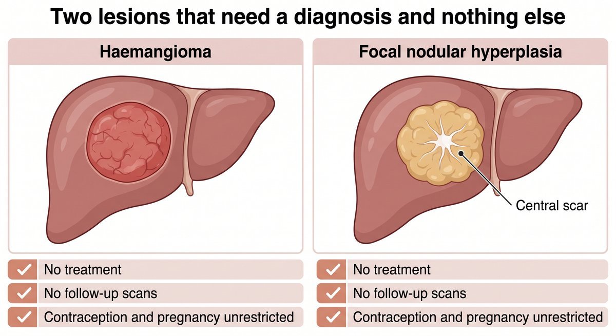
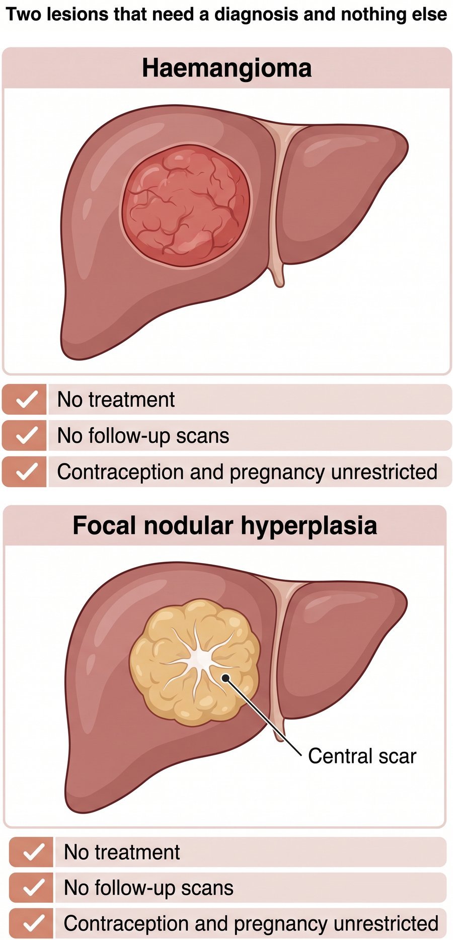
Size does not change this. “Giant haemangioma” describes a measurement, not an indication to operate. Only three things take a haemangioma to a specialist meeting: a coagulopathy in which the lesion interferes with clotting, growth documented across scans, or genuine symptoms from something being compressed.
Focal nodular hyperplasia is not a tumour at all. It is an overgrowth of normal liver tissue in response to an abnormal blood vessel, and it has no malignant potential and no risk of bleeding. It is typically found in a young or middle-aged woman, and shows a central scar on imaging. Once the diagnosis is secure there is no follow-up and no treatment, and again the contraceptive pill is not restricted. Some units image once at a year where diagnostic confidence was less than complete, and then discharge the patient.
Removing a FNH is reserved for a lesion genuinely causing symptoms, and the honest position is scepticism: a small FNH is usually not the cause of the pain it gets blamed for, and operating on it turns a harmless finding into a real operation.
The hepatocellular adenoma is the lesion on this page with real risks: it can bleed, and some types can turn malignant. What is done about it is decided by three things and not by how it looks — the patient's sex, the size of the lesion, and its molecular subtype. The first step is almost never an operation. It is to withdraw the hormonal drive the lesion depends on, and then look again.
| Subtype | How it behaves |
|---|---|
| HNF1A-inactivated | Fat-containing. Low risk of both bleeding and malignant change. One of the two commonest types. |
| Inflammatory | Associated with obesity, metabolic syndrome and alcohol. Carries a bleeding risk. The other of the two commonest. |
| Beta-catenin activated | The highest risk of turning malignant. Removed whatever its size. A much smaller group. |
| Sonic hedgehog activated | A small group, and it carries a bleeding risk. |
| Unclassified | Managed on size and sex, because the subtype is not available to guide it. |
Treatment begins with what is not surgery: stopping the combined contraceptive pill and any anabolic steroid, losing weight, treating the metabolic problem underneath, and stopping alcohol. The liver is re-imaged with MRI at around six months. Many adenomas shrink once the hormonal stimulus is gone, and a shrinking lesion does not need an operation.
An adenoma is removed when any one of the following is true, and the first of them is the one most often missed:
A woman with a stable, low-risk adenoma under 5 cm after hormonal withdrawal is watched rather than operated on, with MRI and a written plan stating what would change the course. Pregnancy needs its own conversation, because adenomas can grow under hormonal stimulation: a woman with an adenoma larger than 5 cm is counselled before she conceives, and an established pregnancy is planned individually with close imaging. Where there are ten or more adenomas, the largest and highest-risk are dealt with; the number alone does not call for more aggressive treatment.
If an adenoma bleeds, the order matters. The bleeding is stopped first by blocking the artery feeding it, and the lesion is removed later, once the patient is stable — emergency removal in an unstable patient is a considerably harder operation with a worse recovery.
Simple liver cysts are extremely common and need no treatment unless they are genuinely causing symptoms. Draining one with a needle is not a treatment — an aspirated cyst always fills again, so it should not be offered as one. A large symptomatic cyst is treated by laparoscopic deroofing: a wide window is cut out of the cyst wall so it cannot re-seal. But before any cyst is opened, the surgeon has to satisfy himself it is not a mucinous cystic neoplasm.
That is the trap on this page. A biliary mucinous cystic neoplasm can look like an ordinary simple cyst on ultrasound. The features that should stop a surgeon are internal septations, nodules on the wall, and thickening or irregularity of the wall — typically in a middle-aged woman.
Deroofing a mucinous cystic neoplasm is the wrong operation. It comes back, and this lesion may already be, or may become, malignant. The correct treatment is complete removal — enucleation, or resecting the segment containing it — not fenestration. A cyst that does not look entirely simple is imaged with MRI and discussed at a joint meeting before anything is done to it.
For that reason the wall of every deroofed cyst goes for examination under the microscope, without exception. If it comes back showing a mucinous lining, the lesion was never a simple cyst and a second, larger operation becomes necessary — which is precisely why the question is asked before the first one.
Polycystic liver disease usually accompanies polycystic kidney disease, and is treated only for symptoms or for a liver enlarged enough to interfere with living. Options run from aspirating and sclerosing a dominant cyst, through fenestration and segmental resection, to transplantation in the severest cases. It is managed jointly with a kidney specialist, because the kidneys usually set the timetable, not the liver.
Every operation here is parenchyma-sparing, because this is benign disease and there is no cancer margin to achieve. That one fact changes the surgery: instead of removing an anatomical section of liver, the lesion is lifted out along its own plane wherever possible, keeping the maximum of working liver. A minimally invasive approach is preferred where feasible, and ultrasound is used inside the abdomen during every liver resection.
| Lesion | The operation, when one is indicated |
|---|---|
| Haemangioma | Enucleation along the compression plane, preferred to anatomical resection — less blood loss and more liver preserved |
| Focal nodular hyperplasia | Parenchyma-sparing resection, or enucleation |
| Hepatocellular adenoma | Parenchyma-sparing resection with a clear margin, laparoscopically where feasible. If it has bled: block the artery first, remove it later |
| Simple cyst | Laparoscopic deroofing with a wide window, and the wall sent for examination |
| Biliary mucinous cystic neoplasm | Complete removal — enucleation or formal resection. Never deroofing |
| Polycystic liver disease | Fenestration, resection or transplantation, jointly with nephrology |
Preparation follows the same routine used before any liver resection — fitness and nutrition built up beforehand, smoking and alcohol stopped at least four weeks ahead, clot prevention and antibiotics at the start of anaesthesia — with one requirement specific to benign disease: an explicit, written statement of why an operation is being advised at all for a lesion that is not cancer. No drain is left after an uncomplicated resection.
Recovery follows a planned day-by-day pathway rather than waiting to see what happens: the breathing tube out and sitting up on the day of surgery, diet and walking from day one, tablets instead of injections for pain by day two, and discharge usually between the third and fifth day after a minor or laparoscopic resection. Going home requires eating, walking, pain controlled on tablets, no fever for a day, and liver function and clotting that are improving. The histology appointment is arranged before discharge — that matters most for a cyst wall and for an adenoma subtype. Two things are reported immediately rather than waited on: bile in the drain with fever, and a rising bilirubin after the third day.
★★★★★
“Dr harsh sir is a good doctor for Liver, Gastro & Cancer Surgeon in Ahmedabad thanks sir. — Milan Pareek”
★★★★★
“I’ve had personal experience with Dr. Harsh Shah. He’s a very knowledgeable and experienced Gastro Surgeon. He’ll explain you all the symptoms patiently and suggest the best route possible at a reasonable cost. He also works towards educating society on the different diseases related to Liver and Pancreas. — Brijesh Nandani”
★★★★★
“We consulted Dr Harsh shah for my Husband as having cyst . surgery was done by Doctor very successfully. today we got discharged. our experience with sir was very good . he is very cooperative, explained about diagnosis very calmly. well Experienced, good Doctor . also thanks to Roshani maam for good cooperation. Thanks sir Dr Priyanka Prajapati — Priyanka Prajapati”
What a benign liver lesion costs depends mostly on whether anything is done to it at all. For a great many patients the entire episode is a consultation and a MRI, and there is no admission. Where an operation is needed, the cost is driven by which operation, whether it is done laparoscopically or open, the room category, the length of stay and what the histology requires — so a figure is given in writing only after the scans have been seen.
Benign liver surgery is covered by health insurance and by CGHS, and the office handles cashless approval and pre-authorisation at Apollo Hospital, Bhat. Approval moves faster when the file states the indication precisely — that the lesion is large and symptomatic, that it has bled or may bleed, or that it is a type carrying a risk of turning. Bring your policy card, photo identification, every scan on disc as well as film, and your current medicine list.
No. A haemangioma, FNH and a simple cyst are not cancers and do not become one. Two lesions here carry a risk that must be managed — a hepatocellular adenoma and a biliary mucinous cystic neoplasm — which is why the lesion is named precisely first.
For a typical haemangioma and a confidently diagnosed FNH, no follow-up imaging is needed at all. Repeat scanning is for an adenoma managed without surgery, and for a mucinous cystic neoplasm that has been removed.
Yes. Neither is contraindicated by a haemangioma or FNH, and the opposite is one of the commonest pieces of wrong advice in this area. An adenoma is different — the pill is stopped, and pregnancy planned with advice first.
No. An aspirated liver cyst refills, so needle drainage is not offered as treatment. A symptomatic cyst is treated by cutting a wide window in its wall laparoscopically, and the wall is sent for examination.
Only for a stated reason: the lesion is large and symptomatic, it has bled or is likely to, or it is a type carrying a risk of turning malignant. If none of those is true, the right treatment is to leave it alone.
Shah’s Gastro, Cancer & Robotic Surgery Centre
Gota, Ahmedabad, Gujarat
Google rating 4.89 from 216 reviews
Patients bring incidental liver lesions to Dr Harsh Shah from across Gujarat — Gandhinagar, Mehsana, Rajkot, Surat, Vadodara and Jamnagar — and from Rajasthan and Madhya Pradesh, often after being told elsewhere that a harmless lesion needs removing. Send the scans on WhatsApp before travelling, so the consultation is spent on the decision.
Bring the MRI, and the scan that first found the lesion. Those two decide whether this needs an operation, a single follow-up scan, or simply an explanation. Call +91-63555-64601 · WhatsApp Dr Harsh Shah's office
Swasth Parivar: twice a month, practical advice on recovery, diet and warning signs. No advertising. Stop any time.
Your number is used for these updates and for the clinic to reach you. It is never sold or passed on. To stop, reply STOP on WhatsApp.