WhatsApp Skip to main content

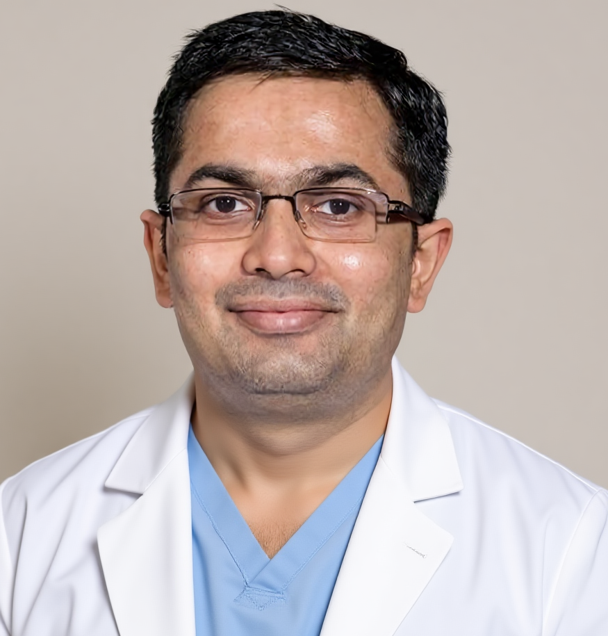
Dr. Harsh J Shah

Fistula MRI: why your surgeon asks for it

Last updated: 18-Oct-2026

Tapping Book on WhatsApp will likely open the WhatsApp app on your phone.

Dr Harsh Shah, GI and proctology surgeon, Ahmedabad

Dr Harsh Shah
MS, MCh (Surgical Gastroenterology), DrNB (Surgical Gastroenterology)
Registered with Gujarat Medical Council, Reg. No. G-38738
GI & Proctology Surgeon
Dr. Jivraj Mehta Hospital, Vasna, Ahmedabad · 18 years of experience
★★★★★ Google rating 4.94 from 18 reviews

In short

An MRI fistulogram is a scan of the area around the anus that maps the fistula tunnel. It shows how the tunnel runs through the control muscles, and any hidden branches or pus pockets. Your surgeon uses it to choose the right operation and to avoid leaving part of the tunnel behind.

What the scan is

It is an MRI of the lower pelvis, taken at special angles along the anal canal. Nothing is pushed into the fistula opening, and there is no X-ray.

The name is a little misleading. An old X-ray test, also called a fistulogram, pushed dye into the opening. That test is no longer used to map a fistula. Today the word means an MRI done with a fistula plan.

The scanner uses a strong magnet and radio waves. The pictures are cut in two special planes: across the anal canal and along it. These show the muscle rings in true shape. A routine pelvic MRI, taken in the usual planes, can miss the tunnel. So the request form should say “MRI fistula protocol” or “MRI fistulogram”.

Some centres also give a dye injection in the arm. It helps tell an active tunnel from old scar. Many scans are clear enough without it.

What the MRI shows

The scan shows the whole path of the tunnel and how much muscle it crosses. It also finds the parts that cannot be felt by a finger.

  • The main tunnel and which type it is: low, high, or curving over the muscle.
  • The inner opening, with its position given as a clock face, such as “6 o’clock”.
  • How much muscle lies above the tunnel. This decides whether cutting is safe.
  • Side branches, including a horseshoe that curves round both sides.
  • Hidden pus pockets, which must be drained first.
  • A second, separate tunnel.
  • Old muscle damage, for example from childbirth or earlier surgery.
  • Signs of Crohn’s disease in the bowel wall.

Why examination alone is not enough

A finger and the eye get the tunnel right only about six times in ten. MRI gets it right about nine times in ten in published studies.

The outer opening on the skin tells little about the path inside. Two tunnels can look the same from outside, yet one is low and simple while the other climbs high through the muscle.

In one large study, the MRI added something important in nearly half of patients. It found extra branches, pus pockets or a high part. One in three fistulas that seemed simple on examination turned out to be complex.

For a fistula that has come back, the difference is larger still. In a well-known trial, when surgeons followed the MRI map, repeat returns fell from more than half to about one in six. Most returns happen at the part the scan had shown and the operation had missed.

When Dr Shah asks for it

He asks for an MRI whenever the tunnel may be more than a simple, low one. A clearly superficial tunnel can go straight to the operation.

When an MRI is usually advised
SituationMRI advised?
Fistula that has come back after surgeryYes, always
More than one opening on the skinYes
Tunnel at the front, in a womanYes
Deep, hard swelling, or pain high in the back passageYes
Weak control, or a difficult birth in the pastYes
Crohn’s disease, known or suspectedYes
A seton in place and the next step is unclearOften
A short, superficial tunnel in a man with good controlUsually not needed

Where MRI is not possible, an ultrasound probe placed in the back passage is a fair choice in skilled hands. It is weaker for deep branches.

How to prepare for the scan

Preparation is simple. Most people need no enema and no special diet. Tell the scan centre about metal in your body and any fear of closed spaces.

  1. Tell the centre about metal. A pacemaker, some heart valves, ear implants and metal fragments can make MRI unsafe. Joint replacements are usually fine.
  2. Mention kidney disease or pregnancy if a dye injection is planned.
  3. Follow the centre’s eating advice. Some ask for a few hours without food when dye is used. Others need nothing.
  4. Leave metal at home. Jewellery, watches, hair clips and belts come off before the scan.
  5. Keep a seton in place. It does not need removing. It even helps the radiologist follow the tunnel.
  6. Expect noise and stillness. You lie on your back for about 20 to 40 minutes. Earplugs are given. Say so in advance if closed spaces worry you.
  7. Bring earlier scans and operation notes. They help the radiologist compare.

Ask for the images on a CD or a link, not only the printed report. Dr Shah reads the images himself, and brings them up in theatre.

When the MRI changes the plan

The scan often decides the operation. It tells the surgeon whether the tunnel can be laid open safely, or whether a seton and a second step are wiser.

What the MRI finds, and what changes
MRI findingWhat it means for the plan
Low tunnel, little muscle crossedLaid open in one operation
High tunnel through much of the muscleLoose seton first, second step weeks later
Hidden pus pocketDrained at the same sitting, before anything else
Horseshoe branchThe deep space is drained and the branches treated, not cut open
A second tunnelBoth tunnels treated in the same sitting
Signs of Crohn’s diseaseJoint care with a gastroenterologist, seton and medicines
Path still unclearLoose seton, then a repeat scan

The final check happens in theatre, under anaesthesia, where the MRI map is matched by feel. How each operation works is explained on the fistula treatment page.

What moves the cost

The cost of the scan depends on the centre, whether a dye injection is used, and whether it is done as an outpatient or during a hospital stay. Many insurance policies cover the MRI when it is part of a planned admission. An outpatient scan may not be covered, so check your policy. For an overview of all four common back-passage problems, see our piles, fissure and fistula clinic page.

Frequently asked questions

Is an MRI fistulogram painful?

No. You lie still on your back while the scanner takes pictures. Nothing is pushed into the fistula opening. The scanner is noisy, and earplugs are given.

Do I need an enema before the MRI?

Usually not. Most centres need no bowel preparation. Some ask for a few hours without food if a dye injection is planned.

Can I have the MRI with a seton in place?

Yes. A loose seton is safe in the scanner and helps the radiologist follow the tunnel.

Is a normal pelvic MRI good enough?

Often not. A fistula needs special angles along the anal canal. Ask for an MRI with a fistula protocol.

Why does Dr Shah want the images and not just the report?

The images show the muscle above the tunnel and any branches in detail. He uses them to plan the operation and brings them into theatre.

Do I need a new MRI if the fistula comes back?

Yes. A repeat fistula needs a fresh map, because the missed branch or opening is what the new operation must find.

Where Dr Harsh Shah sees patients and operates

Dr. Jivraj Mehta Hospital, Vasna, Ahmedabad

Dr. Jivraj Mehta Hospital, Dr Jivraj Mehta Marg, Vasna, Ahmedabad 380007.

Examination under anaesthesia, fistulotomy and loose seton, with a short stay.

+91-63555-64601 · Directions to Dr. Jivraj Mehta Hospital, Vasna

Been asked to get an MRI for your fistula, or already have one? Bring the scan and its report to the consultation, so Dr Shah can see the images during the visit. The plan is made after examination. Call +91-63555-64601 Send the MRI before your visit

Tapping Book on WhatsApp will likely open the WhatsApp app on your phone.

Get health updates from Dr Harsh Shah on WhatsApp

Swasth Parivar: every week, practical advice on recovery, diet and warning signs. No advertising. Stop any time.

Your number is used for these updates and for the clinic to reach you. It is never sold or passed on. To stop, reply STOP on WhatsApp.

Dr. Harsh J Shah
Dr Harsh Shah, Gastro, Cancer & Laparoscopic Surgeon, Ahmedabad
Privacy Overview

This website uses cookies so that we can provide you with the best user experience possible. Cookie information is stored in your browser and performs functions such as recognising you when you return to our website and helping our team to understand which sections of the website you find most interesting and useful.