Skip to main content
Last updated: 21-Nov-2026
Tapping Book on WhatsApp will likely open the WhatsApp app on your phone.
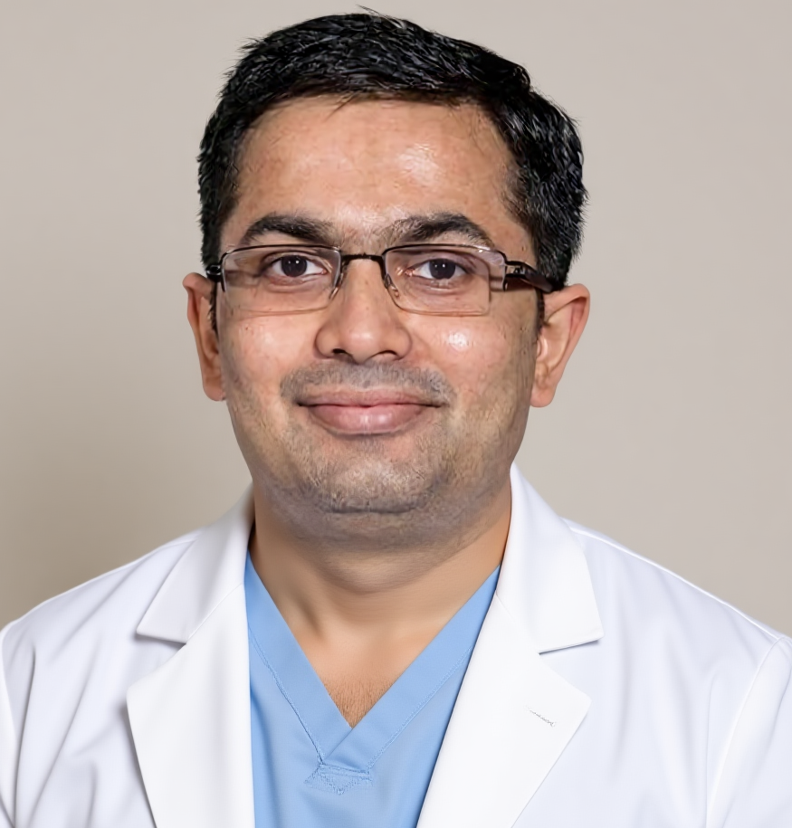
Dr Harsh Shah
MS, MCh (Surgical Gastroenterology), DrNB (Surgical Gastroenterology)
Registered with Gujarat Medical Council, Reg. No. G-38738
Liver & Gastro Surgeon
Dr. Jivraj Mehta Hospital, Vasna, Ahmedabad · 18 years of experience
In short
Budd-Chiari syndrome is a block in the veins that drain the liver. It shows as belly fluid, a tender big liver and pain, and it is often called cirrhosis by mistake.
A Doppler ultrasound is the first test and is often enough. A CT or MR scan then maps the block to plan treatment.
A liver biopsy is rarely needed. Sudden confusion, yellow eyes or fast-worsening illness is an emergency.
For the full picture of this condition, from causes to every treatment choice, read our Budd-Chiari syndrome treatment page. This post answers one question only: how the illness shows itself, and how it is found.
Blood leaves the liver through three large veins. When they are blocked, blood backs up inside the liver, pressure rises and fluid leaks into the belly.
Think of a kitchen sink with a blocked drain. The tap still runs, but the water cannot leave. In the liver, the same back-pressure swells the organ and pushes fluid out into the belly. This is Budd-Chiari syndrome.
How it looks depends on two things: how fast the vein closes, and whether the body has had time to build side channels around the block. A slow block gives the body time to adapt. A sudden block does not.
| Pattern | How fast | What you notice | What it means |
|---|---|---|---|
| Sudden (fulminant) | Days | Confusion, yellow eyes, bleeding tendency, fast belly swelling | An emergency; needs a transplant centre at once |
| Acute | Weeks | Painful, tender liver and fast, tense belly fluid | Liver is congested but not yet scarred |
| Subacute | Weeks to months | Slowly growing belly fluid and vague upper right belly ache | The usual pattern; side channels are forming |
| Chronic | Months to years | Swollen spleen, swollen food-pipe veins, liver that looks cirrhotic | Scarring has begun; easily mistaken for cirrhosis |
Belly fluid, an enlarged tender liver and upper right belly pain are the classic signs. But few people have all three, so the picture is easy to miss.
The textbook trio of belly pain, fluid and a big liver is neither sensitive nor specific. Reading each sign as a pointer works better.
The direction of the belly-wall veins is a useful bedside clue. Veins radiating from the navel point to pressure in the liver’s own vein. Large veins on the flanks and back, carrying blood upward, point to a block in the big vein of the body (the IVC). That block may be a short web, which can often be opened with a balloon.
In India, a slowly growing block in the big vein with leg swelling and visible back veins is a common pattern. It deserves a direct look at that vein.
Both give belly fluid and a stiff, swollen liver. The error happens when nobody asks why the liver veins are blocked.
A chronic block scars the liver, so the scan can look like cirrhosis. If the label “cirrhosis” is accepted without checking the veins, a treatable problem is missed. This matters most in a younger person with no clear cause for liver damage.
Two clues should make a doctor stop and ask the question.
Blood counts can look normal even when a hidden blood disorder is the cause. A normal count does not rule out a clotting tendency.
A Doppler ultrasound of the liver and its veins. It is cheap, safe, repeatable and often enough to make the diagnosis.
A skilled operator checks four things.
The ultrasound should also be read with the heart in mind. A heart ultrasound is part of the workup to rule out heart conditions that mimic this picture.
| Doppler finding | What it means |
|---|---|
| Liver veins not seen, or flow absent, reversed or flat | Outflow block, the core finding |
| Clot or web in a liver vein or the big vein | The block is seen directly |
| Comma-shaped or web-like side channels | Long-standing block with side routes |
| Swollen caudate lobe | The liver is compensating |
| Narrowed big vein with abnormal flow | The block sits at the level of the big vein |
They map the block. They show where it sits, how long it is, and whether the blocking material is clot or something else.
Once the ultrasound points to Budd-Chiari syndrome, CT or MR venography answers the questions that decide treatment. They are not done to repeat the diagnosis.
| Question | Why it matters |
|---|---|
| Where is the block: small liver veins, large liver veins or the big vein? | Decides which treatment is possible |
| Is the block short or long? | A short web can be opened with a balloon; a long one may need a bypass |
| Is the blocking material clot or a growth? | A growth is a different problem with a different pathway |
| Is the portal vein also clotted? | This changes how a bypass is planned |
| How does the liver look? | Shows swelling, shrinkage and benign nodules |
The scan often shows a patchy, uneven liver and a swollen caudate lobe. Large benign nodules are common in a long-standing block. They can look like a liver tumour on a scan, so they need careful reading, a blood test and follow-up rather than a quick label.
MR venography shows the veins well and avoids the iodine dye used in CT. It suits people with kidney problems or dye allergy.
Catheter venography, where a tube is passed into the veins to measure pressure, is not a routine test. It is kept for when treatment will be done in the same sitting, such as opening a web with a balloon.
Usually not. The diagnosis is made by scanning the veins. A biopsy has a small role in selected people.
If a biopsy is done for another reason, it shows congestion around the small central veins and swollen blood channels in the liver. Later, scarring around the veins appears.
A biopsy can help in two situations. The first is when the large veins look open on the scan, but the liver is still congested; the small veins may be the problem. The second is when the amount of scarring will change the choice of treatment. A biopsy that shows nothing does not rule the disease out, because the congestion is patchy.
When the illness worsens fast: confusion, yellow eyes, bleeding tendency or a quick drop in liver function. This needs a transplant centre the same day.
Three questions are asked at the first visit.
A sample of belly fluid is useful on the first visit. It checks protein, rules out infection and sets a baseline before any treatment.
Some settings need extra care. Young women with sudden belly swelling and abnormal liver tests around pregnancy or after delivery should have the liver veins checked, rather than being told it is a pregnancy complication. A clotting disorder behind the block still needs to be looked for later.
Cirrhosis, heart failure, a tight sac around the heart and a clot in the portal vein can all cause belly fluid and a swollen liver.
| Look-alike | How it is told apart |
|---|---|
| Cirrhosis from another cause | Liver veins are open on Doppler; the fluid usually has low protein |
| Heart failure or a tight sac around the heart | Liver veins and the big vein are wide and open; a heart ultrasound shows the cause |
| Portal vein clot | The inflow vein is blocked, not the outflow; both can occur together |
| Small-vein blockage after some drugs or herbal teas | Large liver veins are open; a history of the exposure helps |
The two heart look-alikes are the classic trap. They also swell the liver and fill the belly, but the veins are wide, not blocked. A heart ultrasound is the quick way to tell them apart from Budd-Chiari.
Get a Doppler ultrasound of the liver veins before accepting a label of cirrhosis. If it shows a block, see a liver surgeon for mapping.
For what happens once the block is mapped, read our Budd-Chiari syndrome treatment page. If the real cause is high pressure in the portal vein, see our portal hypertension treatment page.
Belly swelling from fluid, a tender or enlarged liver and pain in the upper right belly are the usual first signs. Some people have only mild symptoms, and some have leg swelling too.
A Doppler ultrasound of the liver veins is the first test. It shows whether blood is flowing in the veins, and it is often enough to make the diagnosis.
Both cause fluid in the belly and a hard, swollen liver. If nobody checks whether the liver veins are open, the problem is called cirrhosis and the real cause is missed.
Usually not. The diagnosis rests on scans of the veins. A biopsy is kept for special situations, such as when the large veins look normal but the liver is still congested.
These scans map where the block is and how long it is. That decides whether a simple balloon opening is possible or a different treatment is needed.
Yes. In rare cases it causes sudden liver failure with confusion and yellow eyes. That is an emergency and needs a transplant centre at once.
Yes. Weak heart pumping or a tight sac around the heart can swell the liver and belly in the same way. A heart ultrasound is part of the workup to rule these out.
For causes and every treatment step, start at the Budd-Chiari syndrome treatment page. For another liver-vein pressure problem, read about portal hypertension. For questions before you travel, call +91-63555-64601.
Dr Harsh Shah — Dr. Jivraj Mehta Hospital, Vasna, Ahmedabad
Dr. Jivraj Mehta Hospital, Dr Jivraj Mehta Marg, Vasna, Ahmedabad 380007.
Consultations, review of reports and scans, operations and follow-up. OPD Monday to Saturday, 9 am to 5 pm.
Appointments: +91-63555-64601 · +91-63590-11009 · Hospital 079-2663 6363
Directions to the hospital · WhatsApp +91-63555-64601
Has your belly been filling with fluid, and has your liver been called enlarged, with no clear reason? Dr Harsh Shah, a liver and gastro surgeon in Ahmedabad, checks first whether the veins that drain your liver are open. If a vein is blocked, the right scans can show where and how long the block is. Bring all your reports, scans and fluid test results. You can also call us or write on WhatsApp. Call For Appointment Book on WhatsApp Tapping Book on WhatsApp will likely open the WhatsApp app on your phone.
Tapping Book on WhatsApp will likely open the WhatsApp app on your phone.
Swasth Parivar: every week, practical advice on recovery, diet and warning signs. No advertising. Stop any time.
Your number is used for these updates and for the clinic to reach you. It is never sold or passed on. To stop, reply STOP on WhatsApp.