WhatsApp Skip to main content

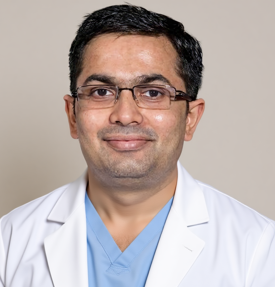
Dr. Harsh J Shah

Infected Pancreatic Necrosis: Why Drain Before Surgery?

Last updated: 20-Nov-2026

Tapping Book on WhatsApp will likely open the WhatsApp app on your phone.

Dr Harsh Shah, gastro and cancer surgeon, Ahmedabad

Dr Harsh Shah
MS, MCh (Surgical Gastroenterology), DrNB (Surgical Gastroenterology)
Registered with Gujarat Medical Council, Reg. No. G-38738
Gastro & Cancer Surgeon
Dr. Jivraj Mehta Hospital, Vasna, Ahmedabad · 18 years of experience

In short

Infected pancreatic necrosis is dead tissue in and around the pancreas that has become infected after a severe attack of pancreatitis. Today it is treated in steps, not with one large operation.

The steps are to wait for the area to firm up, drain it with a tube or a stent, and remove dead tissue only if the drain alone does not settle the infection. Many people need nothing beyond the drain.

Open surgery on a fresh, inflamed abdomen did more harm than good. It is kept as a last resort.

The full plan for an attack of pancreatitis, from the first drip to recovery, is on our acute pancreatitis treatment page. This post explains the step-up approach to infected dead tissue, so the plan you are offered makes sense.

Why is the operation no longer the first step?

Because operating early on dead pancreas made patients sicker. In the first weeks the dead tissue is soft, bleeds easily and is stuck to healthy gland and vessels. Cutting into it causes more damage.

For many years the standard answer to infected necrosis was an open operation as soon as infection was suspected. The surgeon opened the abdomen and cleared out the dead tissue by hand. It was a big operation on a person who was already very weak, and the results were poor.

  • The tissue is not ready. Before about four weeks, dead tissue cannot be told apart from living gland. Pulling it out tears healthy tissue and blood vessels.
  • A second blow to the body. A large operation re-ignites the whole-body inflammation. Lungs and kidneys that were recovering can fail again.
  • Late penalties. An open abdomen leaves more people with new diabetes, hernias at the scar and long fistulae, which are tracks that keep leaking pancreatic juice.

A landmark Dutch trial compared the two approaches. Draining first and escalating only when needed gave fewer serious complications than opening the abdomen at once. Treatment around the world has followed since then.

Does every area of dead pancreas need treatment?

No. Most dead tissue is not infected and slowly settles by itself. We treat the person and the infection, not the size of the scan finding.

This surprises many families. A scan may show a large area of dead pancreas in someone who is eating, walking and has no fever. Such a person needs follow-up and patience, not a procedure. What matters is whether there is infection, whether the person is deteriorating, and whether the collection is blocking something.

When dead pancreatic tissue needs a procedure, and when it does not
SituationUsual plan
Dead tissue, no infection, person is wellWatch, with scans if symptoms change. No procedure, however large it looks
Infection suspected, person is stableAntibiotics and support, and wait for the area to firm up
Infection with the person getting worseDrain, using the gentlest route that works
Firm collection blocking the stomach, intestine or bile ductDrain once it is firm, and only if it is causing trouble
Bleeding or a hole in the bowelUrgent treatment of that problem, which may include an operation

What does step-up care mean?

It means starting with the smallest act that could work and moving to a bigger one only if the smaller one fails. The sequence is wait, drain, then remove dead tissue if needed.

  1. Wait and support. Antibiotics, feeding, and intensive care when needed, while the area firms up.
  2. Drain. A tube through the skin, or a stent through the stomach wall, lets the infected fluid out.
  3. Look at the response. The team gives the drain a fair trial of a few days before calling it a failure.
  4. Make the drain bigger or add a second. A larger tube often clears thicker material.
  5. Remove dead tissue gently. Only if the infection is still not settling, and by keyhole or endoscopy, not a large cut.

Many people stop at step two. Their infection settles, the dead tissue gradually dissolves, and no further procedure is needed. That is the aim of the whole approach.

Why wait about four weeks?

Because after roughly four weeks the body builds a firm wall around the dead tissue. A walled area can be drained safely, and the dead material loosens from living tissue.

Early on, the collection is a soft, shapeless mass that merges into the tissue around it. A tube placed then can slip or bleed, and cannot drain thick material. Once the wall forms, the surgeon has a clear cavity to work in.

Four weeks is a target, not a rule that overrides the patient. If someone is getting worse despite full care before the wall has formed, the team drains earlier by the gentlest route, even though it is harder. The principle is to delay as long as the person safely allows. Recent research on postponed drainage showed that waiting on antibiotics was safe in stable patients, and a good number never needed a drain at all.

Through the skin or through the stomach?

There are two routes, and both work. One goes through the skin of the flank, the other goes through the stomach wall using an endoscope. The choice depends on where the collection lies and who is available to do it.

The two step-up routes compared
PointThrough the skin (surgical route)Through the stomach (endoscopic route)
First stepA tube placed through the flank under scan guidanceA wide stent placed through the stomach wall under ultrasound guidance
If more is neededKeyhole clearing through the same trackA scope passed through the stent to remove dead tissue
Outside woundYes, a tube and later a small cutNone
Risk of a leaking trackHigher, because the tube comes out through the skinLower, because the drainage goes into the stomach
Suits whenThe collection lies along the flank or low in the belly, out of reach of the stomachThe collection sits against the back wall of the stomach
NeedsA radiology team and a surgeonA skilled endoscopy team and the right stents

In big collections, the two are often combined: a stent for the central part and a tube for the part that the scope cannot reach. Large studies found that neither route is clearly safer for survival. The endoscopic route leads to fewer leaking tracks and a shorter stay, when the team and equipment are available.

What if the drain is not enough?

If infection continues, the next step is to remove the dead tissue, but gently. The rule is to take out what is loose and leave what is stuck, because stuck tissue still holds living vessels.

Through the skin route, the surgeon uses the drain track as a guide. A small cut is made over the drain. A camera and long instruments are passed into the cavity. Through the stomach route, a gastroscope goes through the stent and clears the debris under direct vision.

  • Several short sessions. Dead tissue separates over days, so the work is spread out instead of done in one long operation.
  • Washing the cavity. Drains left in place are flushed to carry out the remaining debris.
  • Gas of a safe kind. During endoscopic cleaning, carbon dioxide is used and not air, because air entering a blood vessel can be dangerous.
  • A fair rest between sessions. The person is monitored, fed and strengthened in between.

Open surgery is a last resort. It is used when the gentler routes fail or cannot be used. It is also used when another emergency, such as a hole in the bowel or bleeding that will not stop, needs an operation anyway.

Can antibiotics help us wait?

Yes. Once infection is suspected, antibiotics that reach dead tissue are started. In a stable person they may carry the person through until the wall forms. Antibiotics are not given to prevent infection in dead tissue that is not infected.

Using antibiotics without need breeds resistant germs and fungal infection, which make later treatment harder. Doctors choose an antibiotic that reaches dead tissue. They review it as culture results come back. It is a bridge. It does not replace drainage when the person is getting worse.

What can go wrong with these procedures?

These are serious procedures in people who are already very ill, and each has risks. The team plans for the common ones in advance so that help is on hand at once.

Main risks of treating infected necrosis, and the usual response
ProblemWhy it happensWhat the team does
BleedingA weakened artery in the cavity, or a raw wallCT scan of the vessels, then a blocking procedure by the radiologist before any further drainage
Leaking track after the tubes come outThe main pancreatic duct was cut off by the damageEndoscopic stents inside the duct or the stomach, and long-term follow-up
Stent or tube blocksThick debrisWash, exchange, or add a soft inner tube
Hole in a nearby organThe bowel or stomach is stuck to the cavityDrainage, rest for the bowel, and surgery if needed
Diabetes or poor digestion laterLoss of working glandBlood sugar checks and digestive enzyme capsules with meals

Two rules guide the team. A sudden bleed from a drain or stent is treated as a damaged artery until proven otherwise. It is blocked before the cavity is touched again. A track that keeps leaking is treated as a cut-off duct until proven otherwise. Our post on the disconnected pancreatic duct explains what that means.

Who decides the route?

A team decides, not one doctor. The surgeon, the radiologist and the endoscopist look at the scan together and choose the route that suits the person’s anatomy and condition.

No one specialty sees every option. The team marks which parts of the collection lie against the stomach, along the colon or deep in the pelvis. A plan is made for each part. It is reviewed at every step. Survivors also need long-term follow-up, because the gland may not recover fully. Our post on follow-up after discharge covers that.

Frequently asked questions

Does infected pancreatic necrosis always need an operation?

No. Many people settle with antibiotics and a drain. Dead tissue is removed only if infection continues, and usually by keyhole or endoscopy.

Why not remove the infected tissue at once?

In the first weeks, dead tissue is soft and stuck to living gland and vessels. Removing it then causes bleeding and more damage.

How long do doctors wait before draining?

About four weeks is the aim, so a wall can form. If a person is getting worse sooner, drainage is done earlier by the gentlest route.

Is draining through the stomach better than through the skin?

Both work. The stomach route leaves no outside wound and fewer leaking tracks. It needs a skilled endoscopy team and a collection against the stomach.

Will I need more than one procedure?

Possibly. Dead tissue loosens over days, so cleaning is often done in several short sessions rather than one long operation.

What happens after the cavity heals?

You need regular follow-up. Some people develop diabetes or poor digestion, and a few have a duct problem that causes leaking or new collections.

Related pages

For the whole picture of an attack of pancreatitis, start at the acute pancreatitis treatment page. For questions before you travel, call +91-63555-64601.

Where Dr Harsh Shah sees patients and operates

Dr Harsh Shah — Dr. Jivraj Mehta Hospital, Vasna, Ahmedabad
Dr. Jivraj Mehta Hospital, Dr Jivraj Mehta Marg, Vasna, Ahmedabad 380007.
Consultations, review of reports and scans, operations and follow-up. OPD Monday to Saturday, 9 am to 5 pm.
Appointments: +91-63555-64601 · +91-63590-11009 · Hospital 079-2663 6363
Directions to the hospital · WhatsApp +91-63555-64601

Told that dead tissue in the pancreas is infected and wondering if a big operation is coming? Dr Harsh Shah, a gastro and pancreas surgeon in Ahmedabad, explains why we now drain first and operate only if we must. You can also call us or write on WhatsApp. Call For Appointment Book on WhatsApp Tapping Book on WhatsApp will likely open the WhatsApp app on your phone.

Tapping Book on WhatsApp will likely open the WhatsApp app on your phone.

Get health updates from Dr Harsh Shah on WhatsApp

Swasth Parivar: every week, practical advice on recovery, diet and warning signs. No advertising. Stop any time.

Your number is used for these updates and for the clinic to reach you. It is never sold or passed on. To stop, reply STOP on WhatsApp.

Dr. Harsh J Shah
Dr Harsh Shah, Gastro, Cancer & Laparoscopic Surgeon, Ahmedabad
Privacy Overview

This website uses cookies so that we can provide you with the best user experience possible. Cookie information is stored in your browser and performs functions such as recognising you when you return to our website and helping our team to understand which sections of the website you find most interesting and useful.