Skip to main content
Dr Harsh Shah · Gallbladder Cancer Surgeon · Apollo Hospital, Bhat, Gandhinagar and Gota OPD, Ahmedabad
A gallbladder that is thick-walled, carries a mass or has a large polyp is investigated before it is removed, not after. Where cancer is suspected beforehand the correct operation is not an ordinary keyhole removal but a radical cholecystectomy — the gallbladder taken with the liver bed it sits on and the lymph nodes along the bile duct and the vessels of the liver. Dr Harsh Shah does this at Shah’s Gastro, Cancer & Robotic Surgery Centre and at Apollo Hospital, Bhat.
Shah’s Gastro, Cancer & Robotic Surgery Centre
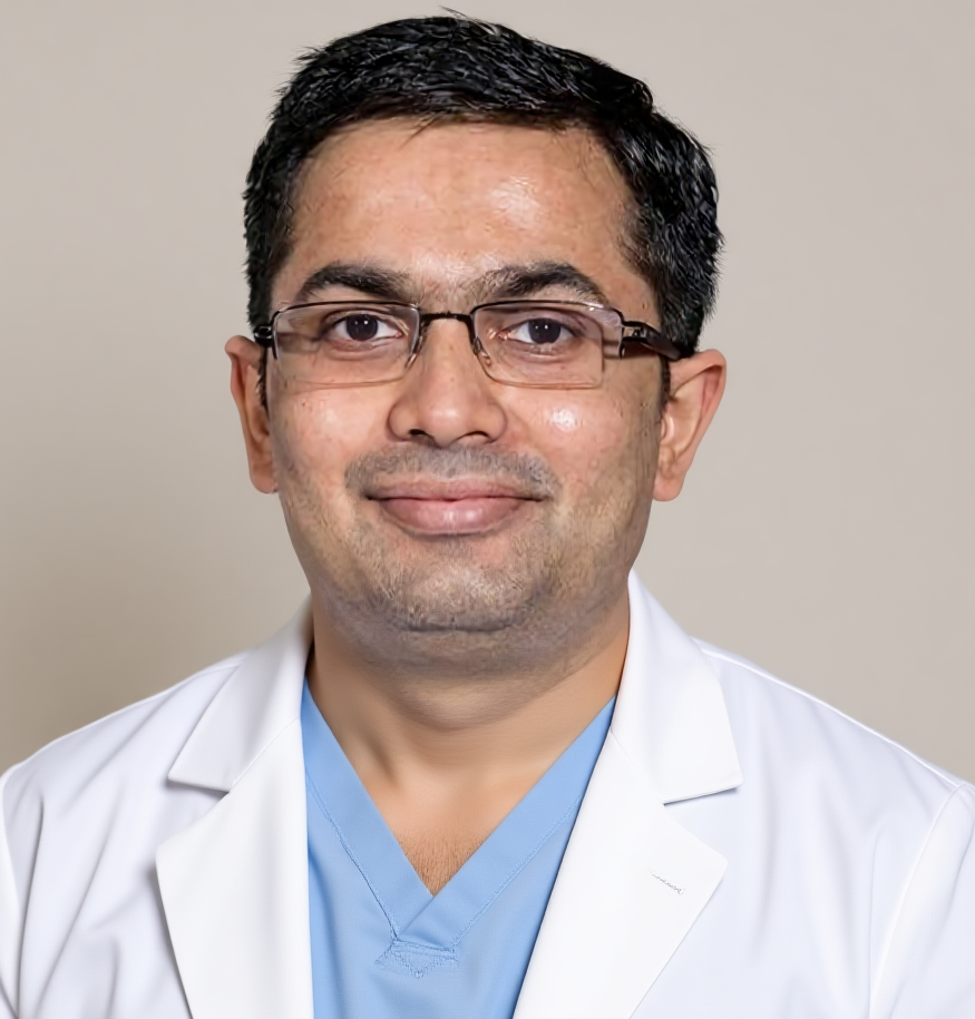
Dr Harsh Shah
MS, MCh (Surgical Gastroenterology), DrNB (Surgical Gastroenterology)
Gallbladder Cancer Surgeon
· Apollo Hospital, Bhat, Gandhinagar · Gota OPD, Ahmedabad
Qualified twice over in surgical gastroenterology and trained in high-volume gastrointestinal cancer units, Dr Harsh Shah takes the suspicious gallbladder as routine work — the thickened wall, the polyp that will not settle, and the cancer that must come out with the liver bed and the nodes. Google rating 4.89 from 216 reviews at Gota, 4.94 from 17 at Apollo Bhat.
Gallbladder cancer is far commoner in India than in the countries whose guidelines are usually quoted, and commoner in women. A wall that is described as thickened is therefore investigated rather than filed as chronic cholecystitis.
Most gallbladders removed here come out for stones, and most thick walls are the scar of years of inflammation. But gallbladder cancer is unusually common across northern and western India and commoner in women. A report reading chronic cholecystitis, wall thickening describes what the ultrasound saw; it is not a verdict on what it means.
What raises the question: focal or uneven thickening rather than even all the way round; a lump in the wall or filling the gallbladder; a polyp of ten millimetres or more, broadly based, growing between scans, or in a patient over fifty; a wall turned to calcium, the porcelain gallbladder, itself a reason to remove it; weight loss, a dull ache under the right ribs, itching or yellow eyes. The operation for a stone gallbladder and the operation for a cancerous one are not the same, and the choice cannot be made afterwards.
Ultrasound raises the question; a multiphasic CT and an MRI with MRCP answer it. A biopsy is not needed before removing a clearly resectable mass, and a negative biopsy never closes the question.
Multiphasic CT of the chest, abdomen and pelvis. Contrast timed in phases, so the liver next to the gallbladder, the nodes along the bile duct and the vessels entering the liver are each seen separately. This is the study that shows whether disease has spread and whether the liver can be cleared with the gallbladder.
MRI with MRCP. A map of the bile ducts made without putting anything into them. Where the tumour sits near the neck of the gallbladder or the junction of the ducts, MRCP shows how far along the duct the disease runs — and that length, not the size of the lump, decides how much has to be removed.
CA 19-9 and CEA. A baseline to compare against later; neither makes nor excludes the diagnosis. CA 19-9 cannot be interpreted when the bilirubin is high or the bile ducts are infected — it is repeated after the jaundice is relieved rather than acted on as it stands.
Staging laparoscopy. Part of the plan, not a failure of the scans: a camera goes in first under the same anaesthetic, because small peritoneal deposits are common here and are what cross-sectional imaging misses most often. Blood count, kidney and liver function, albumin and clotting are checked in the same window; they decide fitness, and cholestasis interferes with clotting.
About biopsy. Tissue is required before chemotherapy, not before removing a mass the scans show can be removed. Brushings miss often enough that a negative result never excludes cancer, and needling risks seeding the track.
Several benign conditions look exactly like gallbladder cancer on a scan. They are separated not by arguing about the images but by planning an operation that is correct whichever it turns out to be.
The commonest imitator is xanthogranulomatous cholecystitis — a fiercely inflamed gallbladder whose wall thickens, whose planes against the liver disappear and whose nodes enlarge. On CT it can be indistinguishable from cancer, the distinction often made only under the microscope. Adenomyomatosis produces focal thickening, a gallbladder shrunken onto stones can look like a tumour, and a benign growth of the gallbladder or adjacent duct occasionally behaves exactly like a malignant one.
Because they are sometimes not separable before surgery, the plan has to be safe both ways: the gallbladder is not squeezed, punctured or torn, it comes out in a retrieval bag, it is opened and inspected in theatre, and it goes to the pathologist whatever it looked like. A cancer in an intact specimen remains treatable; one spilled across the abdomen is far worse.
Dr Harsh Shah explains what gallbladder cancer is, who is at risk, and how it is treated.
A radical cholecystectomy removes the gallbladder with the liver it is stuck to and the nodes along the bile duct, hepatic artery and portal vein. An ordinary keyhole cholecystectomy takes the gallbladder alone and leaves both behind.
Where the gallbladder lies against the liver it has no serosa — no membrane between it and liver tissue. That is why a cancer of its wall reaches liver first, and why removing the gallbladder alone leaves disease behind in a predictable place.
The operation therefore takes three things as one specimen: the gallbladder undisturbed; the liver bed it sits on — segments IVb and V, or a formal liver resection where the tumour demands more; and the nodes of the hepatoduodenal ligament, at the cystic duct, along the bile duct and hepatic artery and behind the portal vein. At least six nodes are aimed for, since disease cannot be called node-negative from a handful, and node status is what the adjuvant decision rests on.
Two things are deliberately not routine. The bile duct comes out only when the cystic duct margin is involved on frozen section; taking it routinely adds a join between duct and intestine, and a lifelong risk of bile duct infection, without improving survival. And port sites are no longer cut out, having been shown not to change outcome. Where disease is confined to the innermost lining and the gallbladder came out complete, that removal alone is enough.
Cancer reported after a routine removal is common in India. Except for the earliest disease, the answer is re-staging and a second, radical operation.
Because every gallbladder goes to the pathologist, cancer is sometimes reported days after an operation done for what everyone believed were stones. The report is read above all for how deep the tumour went: anything deeper than the innermost lining needs the liver bed and the nodes the first operation did not take.
The scans are repeated first: some patients declare disease elsewhere in those weeks, and re-staging prevents a major resection in someone it cannot help. Where disease is still confined, radical resection follows, generally four to eight weeks after the index operation. It is a hard conversation — a completed operation was not the right one — and it is explained rather than announced.
Where there are deposits beyond the gallbladder, disease on the lining of the abdomen, distant nodes or vessels extensively involved, surgery does not help. Treatment begins with drugs, and with relief of jaundice.
Not every gallbladder cancer is operable, and pretending otherwise costs a patient the time they have. Distant deposits, peritoneal disease, nodes around the coeliac axis or behind the aorta, or tumour encasing the vessels of the liver all put resection out of reach — a judgement made at the multidisciplinary meeting, on the imaging and the findings at laparoscopy.
What follows is real treatment, not a consolation. First-line therapy for advanced biliary tract cancer is now chemotherapy with immunotherapy — cisplatin and gemcitabine plus durvalumab or pembrolizumab — not chemotherapy alone, and the change is recent enough that older advice still circulates. Molecular profiling is sent early, because a meaningful minority carry a change for which a targeted drug exists; the local limit is time and cost, not evidence. Where the ducts are blocked, relieving the blockage comes first.
Out of bed and on clear fluids the day of surgery, eating and walking the next day, home in under a week after an uncomplicated resection. Blood tests around day five say whether the liver is behaving.
Recovery runs on an enhanced recovery pathway rather than on waiting: out of bed and on clear fluids the same day; diet built up, catheter out and walking begun on the first day; liver function and clotting checked daily; and any drain inspected each day for bile.
Around the fifth day the bilirubin and the clotting time are the numbers that matter: both improving is expected, and both rising together after the third day says the liver remnant is struggling and changes the plan that day. Most patients are home within five days of an uncomplicated resection — longer after a larger liver resection or a bile duct reconstruction. Discharge also needs eating, independent walking, a day without fever, and no bile in the drain.
The complications that matter are bleeding needing transfusion, infection, a bile leak, a collection inside the abdomen and, after a large liver resection, the liver not coping. The numbers are given individually at the consent discussion, for the operation actually planned.
Published rates are pooled averages across very different operations, and quoting them here would mislead: a radical cholecystectomy and an extended liver resection are not in the same range. What is written here is what can happen; how likely is discussed in person, for the operation actually proposed, by Dr Harsh Shah himself.
Alongside the general risks of major abdominal surgery — bleeding needing transfusion, wound infection, clots in the legs or lungs, strain on the heart or lungs, and anaesthesia itself — the risks particular to this operation are bile leaking from the cut liver surface or from a join between duct and intestine; a collection of pus in the abdomen; bleeding afterwards; clotting of the portal vein or hepatic artery; fluid in the abdomen or around the lung; a return to theatre; and, after a large liver resection, post-hepatectomy liver failure, the leading cause of death after this class of surgery. Where the duct has been joined to intestine, bile duct infection can recur for life and a narrowing of that join years later is a recognised late problem.
Two things are said before the operation, not after. It may be abandoned if deposits are found at laparoscopy or on opening; that decision protects the patient. And the true stage and margins are known only once the pathologist has finished.
After a complete resection, six months of capecitabine tablets follows, started within eight to twelve weeks. Radiotherapy is added selectively, and only as a multidisciplinary decision.
The histology report answers what the scans could not: how deep the tumour went, whether every margin was cleared, how many nodes came out and how many held disease. Those answers, not the operation, decide what comes next.
For resected biliary tract cancer of every subtype, six months of capecitabine is the established adjuvant treatment, started within eight to twelve weeks rather than left until recovery feels complete. Where a margin was involved or nodes positive, chemoradiation is considered — a selective multidisciplinary decision, not a default.
Review at two weeks for the histology, then three-monthly for two years and six-monthly to five years, with CA 19-9, liver function and scans. Every patient is taught to present the same day with fever and jaundice.
The first visit, about two weeks after going home, checks the wound, goes through the histology line by line, confirms the adjuvant referral and reviews nutrition. Review is then three-monthly for two years and six-monthly to five years — symptoms, weight, CA 19-9 and liver function each time, with scans every three to six months at first and MRCP where a narrowing has to be told from a recurrence.
The instruction that matters most is the simplest: a patient whose bile duct has been joined to intestine must know that fever with shivering, or yellow eyes, means bile duct infection and means presenting the same day. That is a drainage problem, not a wait-and-watch one.
A radical cholecystectomy is covered by health insurance and the major TPA panels; the office prepares the pre-authorisation from the imaging, the histology where it exists and the surgical plan. The figure turns on the extent of liver resection, whether the duct is reconstructed, the room category and the stay — so the estimate follows the imaging, not a telephone call.
★★★★★
“Dr. Harsh Shah operated for a gall bladder tumour and the outcome was excellent. Very skilled surgeon, compassionate approach, and clear communication. Truly grateful. Highly recommended.”
★★★★★
“Dr. Harsh Shah operated my mother for gallbladder. The surgery went very well, and she has recovered nicely. We are truly grateful for his expertise and care throughout the treatment. Dr. Harsh Shah is one of the best gallbladder cancer surgeons in Ahmedabad. His approach is very professional, and he ensures that patients and their families feel confident and supported.”
★★★★★
“Dr. Harsh Shah operated on my mother. The surgery was successful and she is doing very well now. He is the best GI cancer surgeon in Ahmedabad. Thank you so much doctor for the excellent care.”
— Sagar Parmar, Google review, Apollo Hospital, Bhat listing
— sohani rana, Google review, Apollo Hospital, Bhat listing
— Radheshyam Soni, Google review, Apollo Hospital, Bhat listing
Usually not — most thick walls are old inflammation from stones. But focal or uneven thickening, a lump, or a polyp of ten millimetres or more is investigated with a multiphasic CT and an MRCP before an operation is planned.
Because there is no membrane between the gallbladder and the liver it lies against. An ordinary removal leaves behind the liver bed and the nodes, which is exactly where gallbladder cancer goes first, and risks tearing the gallbladder and spilling tumour.
The report is read for how deep the tumour went. Confined to the innermost lining, in a gallbladder that came out whole, nothing further is needed. Deeper, the scans are repeated and a second radical operation is advised.
Not before removing a mass the scans show can be removed. Brushings and needle samples miss often enough that a negative result proves nothing, and needling can seed the track. A biopsy is required before chemotherapy, not before resection.
Because small deposits on the lining of the abdomen are common here and are what a CT misses most often. Ten minutes of looking, under the same anaesthetic, prevents a major resection in a patient it could not have helped.
Shah’s Gastro, Cancer & Robotic Surgery Centre
Gota, Ahmedabad, Gujarat
Google rating 4.89 from 216 reviews
Dr Harsh Shah consults at Gota, Ahmedabad and Apollo Hospital, Bhat.
Has a scan described gallbladder wall thickening, a mass or a polyp? Send the ultrasound, the CT or MRI images themselves rather than only the typed report, the liver function tests, and any previous gallbladder operation notes. That combination usually settles whether this needs an operation, and which operation. Call +91-63555-64601 · WhatsApp Dr Harsh Shah's office
Swasth Parivar: twice a month, practical advice on recovery, diet and warning signs. No advertising. Stop any time.
Your number is used for these updates and for the clinic to reach you. It is never sold or passed on. To stop, reply STOP on WhatsApp.