Skip to main content
Dr Harsh Shah · Gastro & Cancer Surgeon · Apollo Hospital, Bhat, Gandhinagar and Gota OPD, Ahmedabad
A choledochal cyst is a bile duct you were born with that is abnormally widened. It is not a cancer, and most people who have one are told exactly that — which is where the danger starts, because a bile duct built this way carries a lifetime risk of turning into bile duct cancer that is far above the ordinary, and that risk climbs with every decade it is left in place. So the operation is not drainage and it is not observation. The abnormal duct is removed completely and the bile is rerouted directly from the liver into the intestine, an operation called a Roux-en-Y hepaticojejunostomy. Dr Harsh Shah performs this at Shah’s Gastro, Cancer & Robotic Surgery Centre, and the two things that decide how well it serves you for the next forty years are how far down the cyst is excised and how the new join is made.
Shah’s Gastro, Cancer & Robotic Surgery Centre
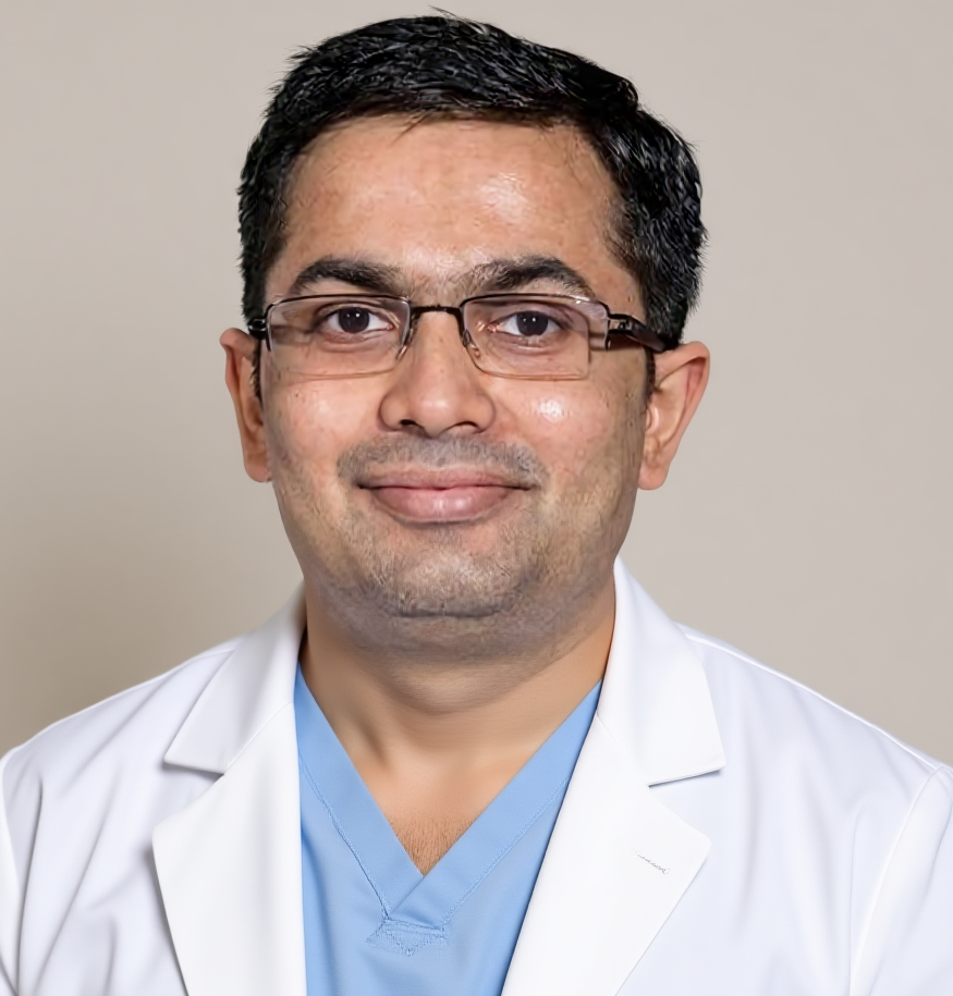
Dr Harsh Shah
MS, MCh (Surgical Gastroenterology), DrNB (Surgical Gastroenterology)
Gastro & Cancer Surgeon
· Apollo Hospital, Bhat, Gandhinagar · Gota OPD, Ahmedabad
Qualified twice over in surgical gastroenterology and trained in high-volume units, Dr Harsh Shah takes on the bile duct work that sits between benign and malignant disease. A choledochal cyst needs a radiologist who reads MRCP properly, a surgeon comfortable dissecting a duct stuck down by years of cholangitis, and a service that will still be calling you in for review a decade later. Google rating 4.89 from 216 reviews at the Gota clinic, and 4.94 from 17 at the Apollo Hospital, Bhat listing.
The bile duct carries bile from the liver to the intestine. In a choledochal cyst that duct is congenitally dilated — sometimes ballooned into a sac, sometimes widened along its whole length. In most people it happens alongside an anomalous pancreaticobiliary ductal junction, a long shared channel that lets pancreatic juice wash back up into the bile duct. That reflux is the presumed reason the lining changes over the years, and the reason this is treated as a premalignant condition rather than a plumbing problem.
Adults rarely present with the textbook childhood picture of pain, jaundice and a lump. What Dr Harsh Shah sees instead is recurrent pain under the right ribs; cholangitis with fever, rigors and yellow eyes; repeated pancreatitis with no gallstones and no alcohol to explain it; stones inside the cyst itself; or, increasingly often, nothing at all — an incidental line on a scan ordered for something else.
There is a harder presentation, and it has to be said plainly. In an adult, a choledochal cyst can arrive with the cancer already in it. A thick wall, a nodule on the inside of the cyst, a mass, enlarged lymph nodes or a markedly raised CA 19-9 mean cholangiocarcinoma until proven otherwise, and that changes the operation entirely. Imaging and sampling come before anyone assumes a benign cyst.
MRCP is the primary investigation, and nothing substitutes for it. It shows the type of cyst, how far it extends into the liver, and whether the bile and pancreatic ducts join abnormally. CT is then done for a different purpose — to look for cancer: wall thickening, a mass, lymph nodes, a shrunken lobe of liver. EUS or ERCP are added where the junction or a possible cancer needs closer definition, and ERCP can also drain the duct when someone arrives with cholangitis.
Bloods at the first visit are LFT, CBC, CRP, clotting, amylase and lipase, and CA 19-9 — with one caveat that is misread constantly. CA 19-9 rises in simple obstruction and in infection, so a high value in a jaundiced or septic patient proves nothing. It is interpreted after the duct has been drained, not before.
In Caroli disease the assessment deliberately goes beyond the liver. Congenital hepatic fibrosis and portal hypertension turn Caroli disease into Caroli syndrome and change the whole plan, so the spleen, the platelet count and the varices are looked for. Caroli disease is also associated with autosomal recessive polycystic kidney disease, which is why renal function and renal imaging are part of the first workup. Assessing only the liver misses half the disease.
Choledochal cysts are grouped by where the dilatation sits, using the Todani classification. It is not academic labelling: the type decides whether the operation is an excision of the duct outside the liver, an endoscopic procedure, a liver resection, or a transplant assessment.
| Type | What is dilated | What is done |
|---|---|---|
| I — the commonest by a wide margin | The bile duct outside the liver, ballooned or widened along its length | Complete excision of the cyst, removal of the gallbladder, and a Roux-en-Y hepaticojejunostomy |
| II | A true outpouching from the side of the duct | The diverticulum is excised and the duct repaired |
| III — choledochocele | The last short segment of duct inside the duodenal wall | Usually endoscopic — sphincterotomy or unroofing. Transduodenal excision if it is large or dysplastic |
| IVa | Both inside and outside the liver | Excision and hepaticojejunostomy, with a liver resection added where the disease inside the liver is confined to one lobe or a segment has shrunk |
| IVb | Several cysts outside the liver | Excision and hepaticojejunostomy |
| V — Caroli disease, confined | Inside the liver only, one segment or lobe | Resection of that part of the liver, which can be curative |
| V — Caroli, widespread, or Caroli syndrome | Throughout the liver, with or without portal hypertension | Assessment for liver transplantation, jointly with hepatology and the transplant service |
For decades the operation was to join the cyst to the duodenum or to a loop of intestine and let it drain, leaving the cyst where it was. That operation is obsolete and is now recognised as harmful. It leaves the abnormal lining in place with its full cancer risk, adds chronic reflux and repeated cholangitis on top, and patients who had it thirty and forty years ago are still presenting today with bile duct cancer.
This matters locally, not just historically. There are people in Gujarat and the neighbouring states, operated on as children, who were told the problem had been fixed. If you or a relative had a “drainage operation” on the bile duct years ago and were then discharged, that is a reason to be reviewed — not reassured. Completion excision is often possible, and where it is not, surveillance has to be intensive rather than occasional.
There is no tablet that shrinks a choledochal cyst, so for type I and type IV the honest answer is that surgery is the treatment. But three situations are managed without cyst excision, and knowing which one applies to you is exactly what the first consultation is for.
A type III choledochocele sits inside the duodenal wall, carries a lower cancer risk than the other types, and is usually dealt with endoscopically rather than by open surgery. Widespread Caroli disease cannot be resected away; it is managed with antibiotics for cholangitis attacks, ursodeoxycholic acid where intrahepatic stones are forming, treatment of portal hypertension, and assessment for transplantation at the right time rather than the last moment. And anyone arriving acutely unwell is not operated on that week: cholangitis is drained and treated first, coagulation and nutrition are corrected, bile is sent for culture, and the definitive operation is done on a prepared patient. Operating through active infection is how a good operation becomes a bad outcome.
For a type I cyst the operation is complete excision of the bile duct outside the liver, removal of the gallbladder as part of the same procedure, and reconstruction with a Roux-en-Y hepaticojejunostomy — a loop of small intestine brought up and joined directly to healthy duct at the liver. Laparoscopic and robotic approaches are appropriate in experienced hands, but the approach is not the point. The quality of the excision and the quality of the join decide the result.
The first decision is how far down the cyst is excised. The duct tapers into the head of the pancreas, and any abnormal lining left behind there keeps its potential to turn into cancer. So the excision goes as far into the intrapancreatic portion as can safely be done. The counter-risk is real — chasing the last few millimetres can injure the pancreatic duct — and balancing those two is a judgement that belongs to a surgeon who does this operation regularly. A long retained distal remnant is the common shortcut, and it is the one that costs the patient later.
The second decision is the join itself. It is made wide, tension-free and mucosa-to-mucosa onto healthy duct. A join made under tension or onto diseased duct narrows over the years, and a narrowed join means recurrent cholangitis, stones forming above it, and repeated procedures. Where dysplasia or cancer is suspected, the margins are checked on frozen section during the operation. The entire specimen goes for histology in every case, because an unsuspected cancer is found in a proportion of adult cysts, and finding it changes what happens next.
The specific hazards Dr Harsh Shah counsels about before this operation are injury to the portal vein or hepatic artery during dissection — the cyst is often densely stuck after years of infection — bile leaking from the new join, an abscess inside the abdomen, and injury to the pancreas or pancreatitis from the lower part of the dissection. Every one of these is discussed by name, with the individual figures for your own case given at the consent discussion rather than quoted on a web page.
After an uncomplicated excision and hepaticojejunostomy most patients sit out of bed the same day, eat as they tolerate from the first day, and go home between the fifth and eighth day. Discharge is not by the calendar: it needs a drain with no bile in it, a falling bilirubin, a normal amylase, and the histology seen and signed off by a named person.
Two things are explained before you leave, and they are the difference between a good operation and a good outcome. The first is cholangitis — fever with rigors and yellowing is an emergency at any point in the future, not something to sleep on. The second is surveillance. A choledochal cyst is not a condition that is treated once and then discharged. Cancer can still arise in the small intrapancreatic remnant, at the join itself, and in the ducts inside the liver. So review continues at three and six months, and then annually for life, with LFT, CA 19-9 and imaging at intervals — looking for narrowing of the join, stones inside the liver, and, most importantly, cancer.
This is planned surgery, so cashless pre-authorisation is completed before admission by the office at Apollo Hospital, Bhat, and CGHS and corporate panels go through the same office. An itemised written estimate is given before you commit. What moves that estimate is the Todani type, whether a liver resection is needed, room category, length of stay, whether ERCP or EUS is done first, and whether you arrive with cholangitis needing drainage before surgery — so a single figure quoted over the phone by anyone, for any patient, is a guess.
★★★★★
“We consulted Dr Harsh shah for my Husband as having cyst . surgery was done by Doctor very successfully. today we got discharged. our experience with sir was very good . he is very cooperative, explained about diagnosis very calmly. well Experienced, good Doctor . also thanks to Roshani maam for good cooperation. Thanks sir Dr Priyanka Prajapati”
★★★★★
“Dr. Harsh Shah operated for a gall bladder tumour and the outcome was excellent. Very skilled surgeon, compassionate approach, and clear communication. Truly grateful. Highly recommended.”
★★★★★
“Dr. Harsh Shah operated my mother for gallbladder. The surgery went very well, and she has recovered nicely. We are truly grateful for his expertise and care throughout the treatment. Dr. Harsh Shah is one of the best gallbladder cancer surgeons in Ahmedabad. His approach is very professional, and he ensures that patients and their families feel confident and supported. Highly recommend him as one of the best cancer surgeons.”
— Priyanka Prajapati, Google review, Apollo Hospital, Bhat listing
— Sagar Parmar, Google review, Gota clinic listing
— sohani rana, Google review, Gota clinic listing
No. A choledochal cyst is a congenital abnormality of the bile duct, not a cancer. What it carries is a lifetime risk of becoming one that is far above the ordinary, and that risk rises with age. That is the whole reason the duct is removed rather than watched. In an adult, though, cancer is specifically looked for before surgery, because occasionally it is already present when the cyst is found.
Draining a choledochal cyst and leaving it in place was the standard operation decades ago and is now considered harmful. The abnormal lining stays, and with it the cancer risk, while reflux and repeated cholangitis are added on top. If a drainage operation has already been done, the right step is specialist review for completion excision, not reassurance.
Yes, you need to be reviewed. What matters is which operation was done. If the cyst was excised and a hepaticojejunostomy made, you still need lifelong annual surveillance. If the cyst was drained and left behind, you carry the original cancer risk and should be assessed for completion excision. The old operation notes are worth hunting for before the appointment.
Laparoscopic and robotic excision are both described and both appropriate in experienced hands, and the approach is decided case by case — how densely the cyst is stuck, whether a liver resection is needed, and whether cancer is suspected.
No, and that is explained at consent rather than introduced afterwards. Cancer can still arise in the part of the duct inside the pancreas that cannot safely be removed, at the join, and in the ducts inside the liver. Annual review with liver function tests, CA 19-9 and imaging continues for life. Being discharged as though the problem were over, after a technically excellent operation, is being under-served.
Shah’s Gastro, Cancer & Robotic Surgery Centre
Gota, Ahmedabad, Gujarat
Google rating 4.89 from 216 reviews
Dr Harsh Shah consults at two addresses: the clinic at Gota, Ahmedabad, and Apollo Hospital at Bhat, Gandhinagar.
Has a scan reported a dilated bile duct, a choledochal cyst, or Caroli disease? Send the MRCP images themselves, not only the typed report. Call +91-63555-64601 · WhatsApp Dr Harsh Shah's office
Swasth Parivar: twice a month, practical advice on recovery, diet and warning signs. No advertising. Stop any time.
Your number is used for these updates and for the clinic to reach you. It is never sold or passed on. To stop, reply STOP on WhatsApp.