WhatsApp Skip to main content
Dr. Harsh J Shah

Klatskin Tumour: Can This Bile Duct Cancer Be Removed?

Last updated: 21-Nov-2026

Tapping Book on WhatsApp will likely open the WhatsApp app on your phone.

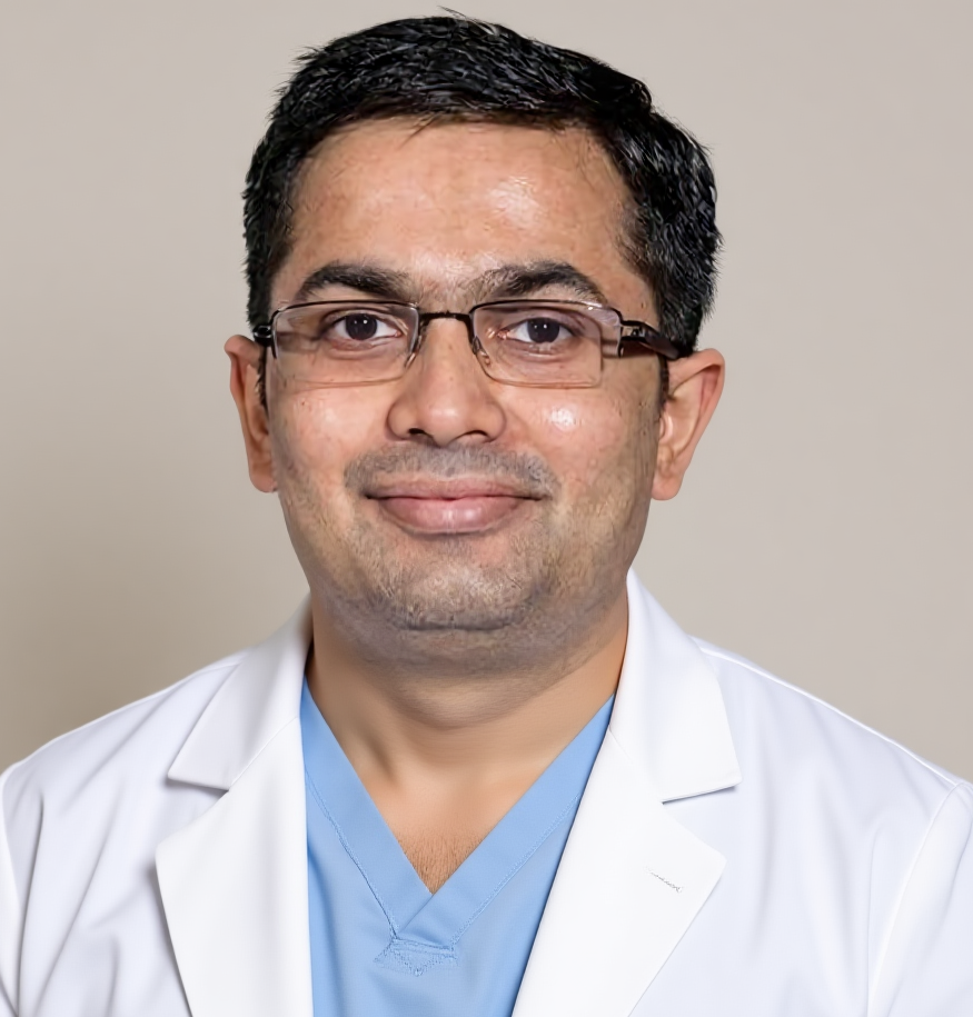
Dr Harsh Shah, liver cancer surgeon, Ahmedabad

Dr Harsh Shah
MS, MCh (Surgical Gastroenterology), DrNB (Surgical Gastroenterology)
Registered with Gujarat Medical Council, Reg. No. G-38738
Liver Cancer Surgeon
Dr. Jivraj Mehta Hospital, Vasna, Ahmedabad · 18 years of experience

In short

A Klatskin tumour sits where the right and left bile ducts leave the liver. Removing it means taking the bile duct and half of the liver together.

Before surgery, only the side of the liver that will stay is drained. The side to be removed is left alone.

If surgery is not possible, chemotherapy with an immune medicine and a stent are used. The scans decide the path.

For the complete picture of this cancer, from tests to surgery and medicines, read our perihilar cholangiocarcinoma treatment page. This post answers one question: how doctors decide whether it can be removed, and how.

What is a perihilar (Klatskin) bile duct cancer?

It is a cancer at the hilum of the liver, the spot where the right and left bile ducts join to form one duct.

Every part of the liver sends bile into small ducts. These join into a right duct and a left duct. The two meet just outside the liver, and the joined duct carries bile to the gut. A cancer here blocks both sides. The skin and eyes turn yellow, urine turns dark, stools turn pale and the skin itches.

The doctors call it perihilar cholangiocarcinoma. Many people know it as a Klatskin tumour. It grows slowly along the ducts and creeps towards the blood vessels that lie right beside them. That closeness is why the operation is big and needs a planned approach.

Which scans show if it can be removed?

An MRI of the bile ducts and a special CT scan. Together they show how far the cancer runs along each duct and which blood vessels it touches.

The surgeon groups the cancer by how high it climbs into the liver. This is called the Bismuth-Corlette type. The type guides which part of the liver must come out.

How the type of Klatskin tumour guides the plan
Where the cancer reachesUsual plan
Below the join of the two ductsBile duct removed, often with a small piece of liver
At the join of the two ductsBile duct removed with the liver part beside it
Into the right ductRight side of the liver removed with the bile duct
Into the left ductLeft side of the liver removed with the bile duct
Into both sides, deep in the liverUsually not removable; chemotherapy first, or a transplant in a few chosen people

Other points also decide the plan. The liver left behind must be big enough to work. The main blood vessels of the liver on both sides must not be wrapped by the cancer. And the cancer must not have spread beyond the liver.

Why drain only one side of the liver?

Because the side that will be removed does not need to be saved. A tube in the side that stays helps it recover and avoids needless infection.

Jaundice in a Klatskin tumour is not always drained before surgery. A stent or tube is used for a clear reason: fever from infection, severe itching, very high bilirubin or a wait of several weeks. When it is needed, the surgeon plans it with the scan.

  • Drain the side that will stay. This lets that part of the liver work well again before the operation.
  • Leave the other side. A tube there adds infection risk and gives no benefit, as it will be removed.
  • Choose the route with the scan. A camera passed through the mouth or a thin tube through the skin can both be used.

Putting stents in both sides without a plan can cause infection and can make the operation harder. So ask the surgeon to see the films before any stent is placed.

What if the liver left behind is too small?

The blood vessel to the side that will be removed can be blocked first. This makes the other side grow over a few weeks, so surgery becomes safe.

Removing half of the liver is safe only when the other half is big enough to do the work. If the scan shows it is small, an expert radiologist blocks the vein feeding the side to be removed. This is called portal vein embolisation. The healthy side then grows. A repeat scan checks the size before the date of surgery is fixed.

People with a liver that was already damaged by an earlier illness need a larger safe margin, so the surgeon checks this more closely.

What is the operation?

The bile duct, half of the liver, the caudate lobe and the nearby lymph glands are removed together. The remaining ducts are joined to a loop of small bowel.

The operation has five parts, done in one sitting.

  • The bile duct outside the liver is removed, with the gallbladder.
  • Half of the liver, right or left, is removed as the scan advises.
  • The caudate lobe, a small part of the liver behind the ducts, is always removed with it, because cancer can hide in its tiny ducts.
  • The lymph glands around the duct and the liver artery are cleared.
  • A new bile channel is made by joining the bile ducts of the remaining liver to a loop of small bowel. This is called hepaticojejunostomy.

If the cancer touches the large vein feeding the liver on one side, that piece of vein can be removed and joined again. A wrapped artery is harder and is handled only in a few cases.

Dr Harsh Shah first looks inside with a small camera to find hidden spread on the liver or belly lining. If none is seen, the main operation is done through an open cut. He does not use a robot. This is a large operation and needs a team that does liver surgery often, with a good intensive care unit.

What does the surgeon check during surgery?

The cut ends of the ducts and the lymph glands. A clean edge is the single most useful sign that all the cancer was taken out.

Cancer can spread along the lining of the duct, further than the scan shows. So each cut duct end goes to the pathologist for a quick look while you are asleep. If cancer cells are found, the surgeon cuts the duct again until the edge is clean. This may need more than one try.

If a gland far from the liver, such as near the main artery behind the belly, holds cancer, the picture changes. The cancer is then no longer local, and the surgeon may stop the big operation to spare you the risk.

What treatment follows the operation?

Chemotherapy tablets of capecitabine for about six months. They lower the chance that the cancer returns.

When the final report is ready, a cancer doctor plans the medicine. Capecitabine is a tablet taken at home in cycles. If the report shows cancer at a cut edge, radiation may be added.

Follow-up after the operation
WhatWhy
Clinic visits with blood tests, including CA 19-9To catch a return early
Scans at planned gapsTo look at the liver and glands
Fever, yellowing or painMay mean a blocked or infected new bile channel, so call us early

A small leak of bile from the new join can happen after surgery. It is watched for in hospital and usually settles with a drain.

What if it cannot be removed?

Chemotherapy with an immune medicine is the main treatment. A stent keeps bile flowing and eases itching.

When the cancer sits deep in both sides of the liver, wraps both main vessels or has spread, an operation does not help. The usual start is two drugs, gemcitabine and cisplatin, with an immune medicine added. Tests on the tumour sometimes find a change that a targeted tablet can act on, so ask for them.

  • A metal stent keeps the bile ducts open. If it blocks, it is changed.
  • Radiation or other local treatment is used for some people whose cancer stays inside the liver.
  • A liver transplant is possible only for a small number of chosen people with an early tumour and no spread to glands. It is done at a few large centres.

If chemotherapy shrinks the cancer a lot, surgery can be reviewed again. Comfort, appetite and itching control matter as much as the scan.

What should you do next?

Collect all scan films, do not allow stents on both sides without a plan, and see a surgeon who does liver and bile duct surgery often.

  • Bring the CT scan and MRI films, not only the reports.
  • Tell us about fever, itching, weight loss and any stent or tube already placed.
  • Ask which side of the liver is to stay and whether it is big enough.
  • Ask who will plan the medicine after surgery, and when it begins.

Frequently asked questions

Can surgery remove a Klatskin tumour for good?

Surgery is the only way to remove it for good, but it is followed by chemotherapy tablets to lower the chance of return. Surgery is possible only when the cancer is confined and the remaining liver is large enough.

Why is half of the liver removed for a bile duct cancer?

The cancer sits where the two liver ducts join and grows along them. Taking the liver part on the involved side, with the duct, is the only way to reach clean edges.

Do I need a stent before the operation?

Not always. A stent is placed for fever from infection, severe itching, very high bilirubin or a long wait. If one is needed, only the side of the liver that will stay is drained.

What is portal vein embolisation?

A radiologist blocks the vein feeding the side of the liver that will be removed. The other side then grows over a few weeks, so it can safely do all the work after surgery.

Why is the caudate lobe removed with the liver?

The caudate lobe drains into the ducts at the hilum, and cancer can hide in its tiny ducts. Leaving it behind raises the risk that cancer stays.

Is a liver transplant an option for a Klatskin tumour?

Only for a few people with a small early tumour and no spread to glands, after a special course of radiation and tablets. It is done at a small number of large centres.

What treatment follows the operation?

Capecitabine tablets for about six months, with regular blood tests and scans. Your cancer doctor sets the dose and watches for side effects.

Related pages

For the full picture of this cancer and its care, start at the perihilar cholangiocarcinoma treatment page. For the wider group of bile duct and gallbladder cancers, see biliary tract cancer treatment. For questions before you travel, call +91-63555-64601.

Where Dr Harsh Shah sees patients and operates

Dr Harsh Shah — Dr. Jivraj Mehta Hospital, Vasna, Ahmedabad
Dr. Jivraj Mehta Hospital, Dr Jivraj Mehta Marg, Vasna, Ahmedabad 380007.
Consultations, review of reports and scans, operations and follow-up. OPD Monday to Saturday, 9 am to 5 pm.
Appointments: +91-63555-64601 · +91-63590-11009 · Hospital 079-2663 6363
Directions to the hospital · WhatsApp +91-63555-64601

Told you have a growth at the gate of the liver, where the bile ducts join? This is one of the hardest cancers to operate on, and the plan depends on the scans. Dr Harsh Shah, a liver and gastro surgeon in Ahmedabad, studies your films himself and says plainly whether the cancer can come out. Bring every report and scan film. You can also call us or write on WhatsApp. Call For Appointment Book on WhatsApp Tapping Book on WhatsApp will likely open the WhatsApp app on your phone.

Tapping Book on WhatsApp will likely open the WhatsApp app on your phone.

Get health updates from Dr Harsh Shah on WhatsApp

Swasth Parivar: every week, practical advice on recovery, diet and warning signs. No advertising. Stop any time.

Your number is used for these updates and for the clinic to reach you. It is never sold or passed on. To stop, reply STOP on WhatsApp.

Dr. Harsh J Shah
Dr Harsh Shah, Gastro, Cancer & Laparoscopic Surgeon, Ahmedabad
Privacy Overview

This website uses cookies so that we can provide you with the best user experience possible. Cookie information is stored in your browser and performs functions such as recognising you when you return to our website and helping our team to understand which sections of the website you find most interesting and useful.