WhatsApp Skip to main content

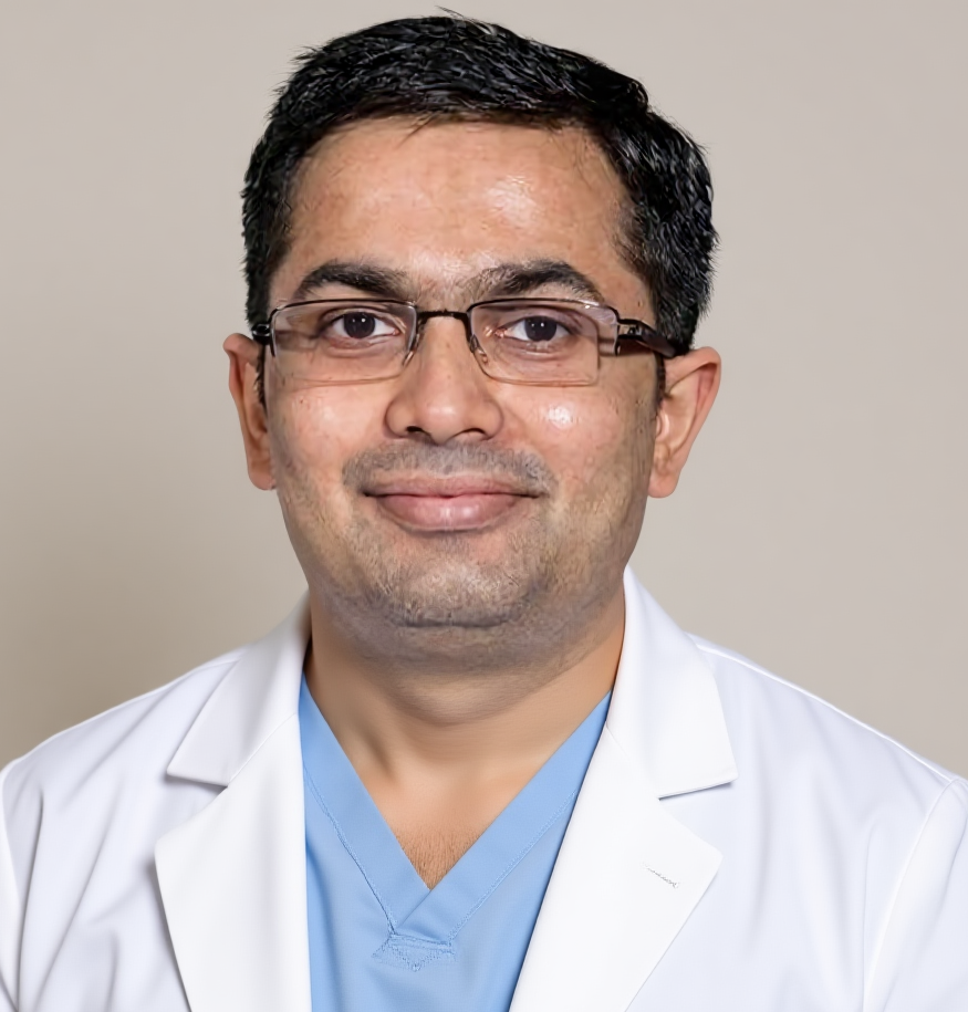
Dr. Harsh J Shah

Fluid Near the Pancreas: What CT and EUS Can Tell You

Last updated: 07-Nov-2026

Tapping Book on WhatsApp will likely open the WhatsApp app on your phone.

Dr Harsh Shah, gastro and cancer surgeon, Ahmedabad

Dr Harsh Shah
MS, MCh (Surgical Gastroenterology), DrNB (Surgical Gastroenterology)
Registered with Gujarat Medical Council, Reg. No. G-38738
Gastro & Cancer Surgeon
Dr. Jivraj Mehta Hospital, Vasna, Ahmedabad · 18 years of experience

In short

After pancreatitis, fluid can gather near the pancreas. A CT scan finds it. A pseudocyst holds fluid alone, while walled-off necrosis also holds dead tissue.

CT shows the size, the wall and the position. EUS looks from inside the stomach and sees solid debris, nearby vessels and the wall more clearly.

Many collections need no procedure. The scans help decide who can simply be watched and who needs drainage.

The full plan for acute pancreatitis, from the first tests to drainage or surgery, is on our acute pancreatitis treatment page. This post covers one step: how CT and EUS are used to read a pancreatic collection.

What is a pancreatic collection?

A pancreatic collection is a pocket of fluid, sometimes with dead tissue, that forms in or around the pancreas after an attack of pancreatitis.

When the pancreas is inflamed, it can leak digestive juice. Some of the gland may also die. The body tries to wall this off, and a pocket forms.

Most pockets are found on a scan done for pain, fever or a poor appetite after pancreatitis. Some are found by chance. A pocket by itself is not always a problem.

Pseudocyst or walled-off necrosis: what is the difference?

Both have a wall, and both form after about four weeks. A pseudocyst holds fluid only. Walled-off necrosis holds fluid and solid dead tissue.

Four kinds of pancreatic collection
NameWhen it appearsWhat is insideWall
Acute fluid collectionIn the first four weeksFluid onlyNo clear wall
Acute necrotic collectionIn the first four weeksFluid and dead tissueNo clear wall
PseudocystAfter four weeksFluid onlyClear wall
Walled-off necrosisAfter four weeksFluid and dead tissueClear wall

The difference matters because the plan differs. Fluid alone often drains easily. Dead tissue is thicker, and it may need more than one step to clear.

What does a CT scan show?

CT is usually the first scan. It shows where the collection lies, how big it is, how thick the wall is, and whether gas is present.

A CT scan with contrast dye also shows whether the pancreas itself is still well supplied with blood. That tells the team how much of the gland has died.

  • Size and position. The scan shows whether the pocket presses on the stomach, bowel or bile duct.
  • Wall. A wall that is fully formed means the pocket is mature and ready for any drainage that may be needed.
  • Gas bubbles. Gas inside the pocket, with fever, can point to infection.
  • Nearby vessels. A CT angiogram can show a swollen blood vessel next to the pocket, called a pseudoaneurysm.

CT has one weakness. Thick fluid and dead tissue can look alike on it. That is why a second test is sometimes needed.

When is MRI or EUS added after a CT?

A second test is added when the CT cannot tell fluid from debris, when the cause of the pocket is unclear, or when drainage is being planned.

MRI, and a special MRI of the bile and pancreatic ducts, picks up solid debris better than CT. It can also show whether the main pancreatic duct has been broken, which affects the treatment choice.

EUS is an endoscopic ultrasound. It gives a close view from inside the stomach, so it adds detail that scans from outside cannot give.

What does EUS add?

EUS sees through the stomach wall to the pocket. It shows solid debris, the wall and any vessel in the path, and it can take a fluid sample.

  • Debris. It shows how much solid material is inside, which separates a pseudocyst from walled-off necrosis.
  • Distance. It measures how close the pocket lies to the stomach wall, which matters if drainage is planned.
  • Vessels. Doppler shows swollen veins or an aneurysm on the route, which the doctor must avoid.
  • Other cysts. When the history is not clear, fluid from the pocket can be tested to see if it is a different kind of cyst.

Our post on EUS explains the test itself, and the post on CT and MRI covers the scans.

How is an EUS done?

You are given sedation, so you sleep through it. A flexible tube goes down through the mouth into the stomach, and the test takes a short time.

  1. You fast for several hours before the test, and your medicines are checked, especially blood thinners.
  2. Sedation is given through a drip in your arm.
  3. The doctor passes the tube through the mouth to the stomach and looks at the pocket with ultrasound.
  4. If needed, a thin needle takes a small fluid sample from the pocket.
  5. You rest for a short time and usually go home the same day, unless a procedure is done as well.

Serious problems from EUS are uncommon. They include bleeding, infection and a sore throat. Your doctor explains the risks for you before the test.

Does every collection need treatment?

No. A small pocket with no symptoms is often only watched with repeat scans. Treatment is for pockets that cause trouble or are infected.

A pocket may need treatment if it blocks the stomach or bile duct, causes steady pain, is infected, or keeps growing. Many pockets shrink without help.

The scans tell us what is inside and how mature the wall is. How the pocket is then drained, by endoscope, by a tube or by an operation, is a separate choice. It is covered on our acute pancreatitis treatment page.

What should you bring to the consultation?

Bring every CT, MRI and ultrasound report with the images on a disc, your discharge card from the pancreatitis admission, and your blood test reports.

  • All CT, MRI and ultrasound images and reports, oldest first
  • The discharge card from the pancreatitis admission
  • Blood reports, especially amylase, lipase and counts
  • A list of your medicines, including blood thinners
  • Notes on pain, fever, vomiting, weight loss and diet

If your attack was linked to gallstones, our post on gallstone pancreatitis explains why the gallbladder matters.

Frequently asked questions

What is the difference between a pseudocyst and walled-off necrosis?

A pseudocyst holds fluid only. Walled-off necrosis holds fluid and solid dead tissue. Both have a clear wall and form after about four weeks.

Is a CT scan enough to diagnose a pancreatic collection?

CT finds the collection and shows its size and wall. It can miss solid debris, so MRI or EUS is added when the answer changes the plan.

Is EUS safe?

Serious problems are uncommon. The test is done under sedation, and the risks, such as bleeding or infection, are explained to you before it.

Do I need to stay in hospital for an EUS?

For the test alone, you usually go home the same day once the sedation wears off. A drainage procedure may need a longer stay.

Will a pseudocyst go away without treatment?

Many small ones do, especially when there are no symptoms. Repeat scans show if the pocket is shrinking or growing.

Can a pancreatic cyst be something other than a pseudocyst?

Yes. When there was no clear attack of pancreatitis, EUS and a fluid test help to separate a pseudocyst from other kinds of cyst.

Which test is needed first?

A CT scan with contrast is usually first. Your doctor then adds MRI or EUS only if it will change what is done.

Related pages

For the whole pathway of acute pancreatitis, start at the acute pancreatitis treatment page. For questions before you travel, call +91-63555-64601.

Where Dr Harsh Shah sees patients and operates

Dr Harsh Shah — Dr. Jivraj Mehta Hospital, Vasna, Ahmedabad
Dr. Jivraj Mehta Hospital, Dr Jivraj Mehta Marg, Vasna, Ahmedabad 380007.
Consultations, review of reports and scans, operations and follow-up. OPD Monday to Saturday, 9 am to 5 pm.
Appointments: +91-63555-64601 · +91-63590-11009 · Hospital 079-2663 6363
Directions to the hospital · WhatsApp +91-63555-64601

Had pancreatitis, and a scan now shows fluid or a "cyst" near the pancreas? Dr Harsh Shah goes through your CT and reports with you, explains what the collection is made of, and says whether a second test such as EUS would help. Book a visit and bring every scan and report you have. You can also call, or message us on WhatsApp. Call +91-63555-64601 Ask on WhatsApp

Tapping Book on WhatsApp will likely open the WhatsApp app on your phone.

Get health updates from Dr Harsh Shah on WhatsApp

Swasth Parivar: every week, practical advice on recovery, diet and warning signs. No advertising. Stop any time.

Your number is used for these updates and for the clinic to reach you. It is never sold or passed on. To stop, reply STOP on WhatsApp.

Dr. Harsh J Shah
Dr Harsh Shah, Gastro, Cancer & Laparoscopic Surgeon, Ahmedabad
Privacy Overview

This website uses cookies so that we can provide you with the best user experience possible. Cookie information is stored in your browser and performs functions such as recognising you when you return to our website and helping our team to understand which sections of the website you find most interesting and useful.