Skip to main contentLast updated: 07-Nov-2026
Tapping Book on WhatsApp will likely open the WhatsApp app on your phone.

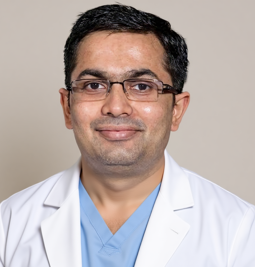
Dr Harsh Shah
MS, MCh (Surgical Gastroenterology), DrNB (Surgical Gastroenterology)
Registered with Gujarat Medical Council, Reg. No. G-38738
Liver Cancer Surgeon
Dr. Jivraj Mehta Hospital, Vasna, Ahmedabad · 18 years of experience
In short
Before liver surgery you need a contrast CT, usually a liver MRI, and tests that check the bowel and your blood.
The scans map every liver deposit, look for cancer elsewhere, and measure how much healthy liver would be left.
Scans taken long ago are often repeated, because the cancer and the liver can change during chemotherapy.
The tests, treatment stages and choices for bowel cancer that has reached the liver are on our colorectal liver metastases page. This post covers one step: the scans and tests done before the liver operation.
Each scan answers a different question. Together they tell the surgeon what can be removed, what must stay, and whether surgery is the right step.
Liver surgery for bowel cancer is planned like a map. The surgeon needs to know where each deposit lies, and how close it sits to the large blood vessels and bile ducts.
The team also has to be sure the cancer is not growing somewhere else. An operation on the liver helps only when all the visible disease can be dealt with.
The core set is a contrast CT of the chest, abdomen and pelvis, a liver MRI, a look at the bowel, and blood tests.
| Test | What it shows | Why it matters |
|---|---|---|
| Contrast CT of chest, abdomen and pelvis | Liver deposits, blood vessels, lungs, lymph nodes | Maps the disease and checks for spread outside the liver |
| Liver MRI with contrast | Small deposits and fine detail inside the liver | Finds deposits CT can miss, so the plan is complete |
| PET-CT scan | Cancer activity in the whole body | Used in selected patients to rule out hidden spread |
| Colonoscopy and bowel imaging | The bowel tumour and the rest of the bowel | Confirms the first tumour is dealt with, and no second one is missed |
| Liver volume measurement | Size of the liver that would be left | Shows how much can be removed safely |
| Blood tests including CEA | Liver function, clotting, tumour marker | Checks fitness for surgery and gives a baseline to follow |
Not every patient needs every test. Your surgeon picks the set that fits your scans, your chemotherapy history and your fitness.
MRI shows soft tissue in finer detail. It can find small deposits that a CT does not clearly show, especially after chemotherapy.
Some MRI scans use a special dye that the healthy liver takes up. The deposits do not, so they stand out against the normal liver.
This matters because every deposit missed on scans is a deposit left behind. A full picture before surgery lowers the chance of a surprise on the table.
A PET-CT is not needed for everyone. It is used when the surgeon wants to check the whole body for hidden cancer before a big operation.
It helps most when the cancer has come back, when the CEA level is rising without a clear cause, or when a scan shows something doubtful outside the liver.
A PET-CT is also less useful soon after chemotherapy. Treatment can quieten small deposits so that they do not light up. Your team will time it with this in mind.
Software on a CT or MRI scan outlines the liver and calculates the size of the part that would remain after surgery.
The remaining liver must be large enough to do the work of the whole organ. It also needs its own blood supply and bile drainage.
How the operation itself is carried out is explained in our post on how surgeons remove bowel cancer from the liver.
Scans are usually repeated during and after chemotherapy, so the surgeon plans from fresh pictures and not from the first ones.
Chemotherapy can shrink deposits, and some may fade on the scan. The surgeon still plans to deal with the place where each deposit was seen.
Surgery is usually planned a few weeks after the last treatment, with a new scan close to the date. Our post on chemotherapy before and after liver surgery explains the timing.
Most scans need an empty stomach for some hours, and a contrast dye through a vein. Tell the staff about allergies and kidney problems.
The surgeon looks at the pictures themselves. A written report alone is not enough to plan a liver operation.
Scans are very good but not perfect. During the operation the surgeon scans the liver directly with an ultrasound probe.
This check can find tiny deposits that were too small to see before. It can also confirm that the planned cut keeps clear of key vessels.
Sometimes the plan changes on the table. You are told about this during consent, so nothing comes as a shock afterwards.
Ask which scans are still needed, whether older ones must be repeated, and how much liver the scans say will be left.
If you are still unsure whether the deposits can be removed at all, read our post on whether colon cancer in the liver can be removed.
A CT is a strong first scan, but it can miss small deposits. A liver MRI adds fine detail, and together they give the surgeon a fuller map.
Not always. It is used in selected patients, for example when the cancer has returned or a scan shows something doubtful outside the liver.
Older scans help for comparison, but the surgeon usually wants a recent set. Cancer and the liver can change during chemotherapy.
It is safe for most people. Tell the staff about allergies, kidney disease or a past reaction, so they can plan around it.
Yes, very small deposits can be missed. This is why the surgeon also checks the liver with ultrasound during the operation.
CEA is a marker that often rises with bowel cancer. It is used as a baseline before surgery and followed afterwards to watch for any return.
Bring the films on a disc as well as the report. The surgeon plans the operation from the pictures themselves, not only from the written words.
For the full picture of bowel cancer in the liver, start at the colorectal liver metastases page. For questions before you travel, call +91-63555-64601.
Dr Harsh Shah — Dr. Jivraj Mehta Hospital, Vasna, Ahmedabad
Dr. Jivraj Mehta Hospital, Dr Jivraj Mehta Marg, Vasna, Ahmedabad 380007.
Consultations, review of reports and scans, operations and follow-up. OPD Monday to Saturday, 9 am to 5 pm.
Appointments: +91-63555-64601 · +91-63590-11009 · Hospital 079-2663 6363
Directions to the hospital · WhatsApp +91-63555-64601
Been asked to get a string of scans before liver surgery, and not sure what each one is for? Dr Harsh Shah goes through your reports and films with you, tells you which tests are still missing, and explains what they mean for the operation. Book a visit and bring every scan, disc and report you have. You can also call, or message us on WhatsApp. Call +91-63555-64601 Ask on WhatsApp
Tapping Book on WhatsApp will likely open the WhatsApp app on your phone.
Swasth Parivar: every week, practical advice on recovery, diet and warning signs. No advertising. Stop any time.
Your number is used for these updates and for the clinic to reach you. It is never sold or passed on. To stop, reply STOP on WhatsApp.