Skip to main content
Shah's Gastro, Cancer & Robotic Surgery Centre · Ahmedabad & Gandhinagar
Mid oesophageal cancer treatment in Ahmedabad · Gota OPD, Ahmedabad · Apollo Hospital, Bhat, Gandhinagar
A cancer in the middle third of the food pipe lies directly behind the windpipe and its main branches, and that one fact changes three things. The windpipe is looked at from inside before any plan is made; treatment usually starts with chemotherapy and radiation rather than with an operation; and when an operation is done, the new join is made in the neck, well above the tumour. At Shah’s Gastro, Cancer & Robotic Surgery Centre each of those decisions is taken in order, by one team, at the tumour board.
The team that repeats the endoscopy, reads the staging scans and presents the case at the tumour board is the same team that operates when an operation is right — and that also says plainly when it is not. A middle-third tumour needs all of those skills, because its treatment is often a sequence rather than a single step.
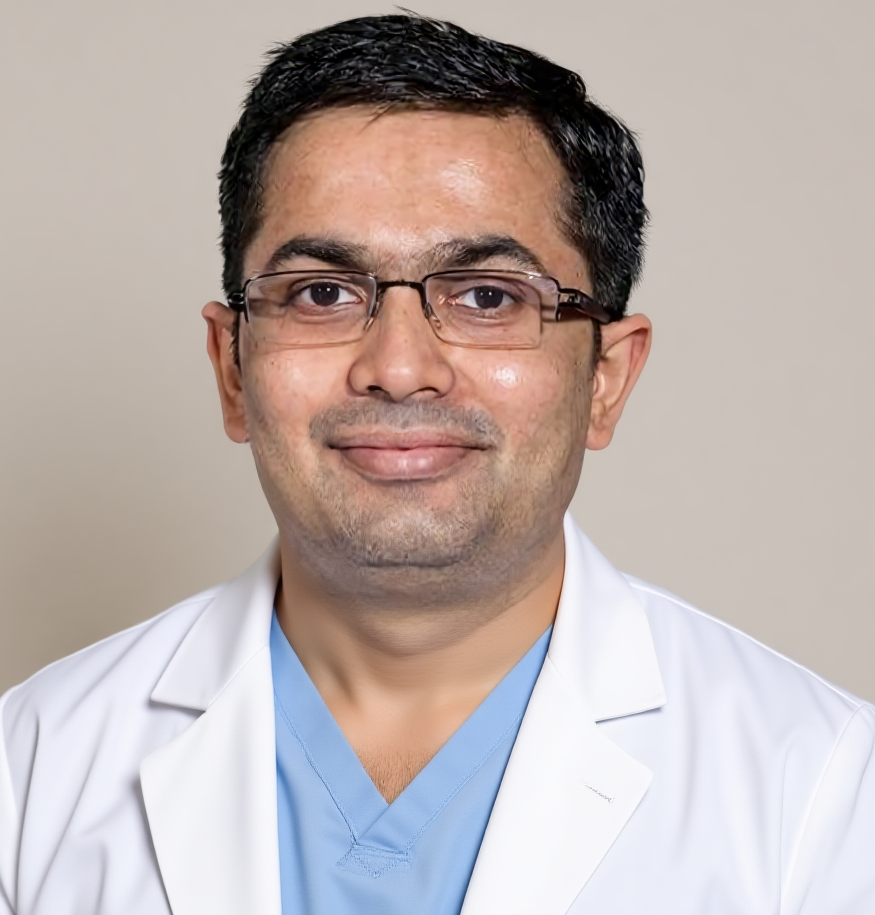
Shah’s Gastro, Cancer & Robotic Surgery Centre. Trained in super-specialty surgical gastroenterology programmes, qualified twice over in the specialty, with 18 years of experience in GI and cancer surgery. Upper GI endoscopy, staging, the tumour board, operations on the food pipe through the abdomen, chest and neck, nutrition support and long-term follow-up sit within one practice.
Dr Harsh Shah
MS, MCh (Surgical Gastroenterology), DrNB (Surgical Gastroenterology)
Registered with Gujarat Medical Council, Reg. No. G-38738
Food Pipe Cancer Surgeon · Apollo Hospital, Bhat, Gandhinagar · Gota OPD, Ahmedabad
Google rating 4.89 from 216 reviews at the Gota clinic, and 4.94 from 17 reviews at Apollo, Bhat.
The food pipe runs from the throat to the stomach in three stretches: the upper part near the neck, the middle part behind the windpipe and the heart, and the lower part that joins the stomach. Endoscopy reports describe a tumour’s position by its distance from the front teeth. When that distance places the growth in the middle stretch, the plan has to account for the airway sitting immediately in front of it, for the type of cancer that the biopsy shows, and for how far above the tumour the surgeon must go to remove it safely.
Three features of a middle-third tumour shape everything that follows: the windpipe and its left main branch lie right in front of it; the biopsy type — squamous cell cancer or adenocarcinoma — decides which treatment comes first; and a safe upper margin usually means the join has to be made in the neck rather than inside the chest.
A tumour that has grown into the wall of the windpipe cannot simply be cut away. A tumour that only presses on it can. Telling the two apart is the first job, and it is done with a bronchoscopy, not guessed from a scan. A late sign such as a cough every time the patient swallows, or a hoarse voice, raises the possibility of an abnormal opening between the food pipe and the airway, and that patient is assessed by the consultant surgeon the same day.
The type of cancer matters as much as the position. Squamous cell cancer and adenocarcinoma follow different treatment pathways, so the biopsy report is read for its type before any treatment is discussed. Squamous cancers of the upper and middle food pipe also travel with a risk of a second cancer in the throat or voice box, which is why a throat examination is part of the work-up for them.
For a middle-third tumour the order is: endoscopy with several biopsies and dye mapping of the edges, a contrast CT of the chest and abdomen, a PET-CT when removal of the cancer is being considered, a bronchoscopy to look at the windpipe from inside, a throat examination for squamous cancer, endoscopic ultrasound only where it changes the decision, and heart and lung tests before any chest operation.
| Check | What it decides for a middle-third tumour |
|---|---|
| Endoscopy with several biopsies, measured distances, photographs, and iodine dye or narrow-band light to map the edges | Cancer or not, its type, and exactly where it begins and ends — the surgeon’s own scope is preferred, because the margin above the tumour is what fixes the operation |
| Contrast CT of the chest, abdomen and pelvis | Spread to the liver, lungs or lymph nodes, and how the tumour relates to the airway |
| PET-CT | Hidden spread that would make a major operation the wrong choice — done for everyone in whom removal is being considered |
| Bronchoscopy | Whether the windpipe or its main branch has been invaded — the single check that most often changes the plan in this part of the food pipe |
| Throat and voice-box examination (for squamous cancer) | A second cancer in the head and neck that would need its own treatment |
| Endoscopic ultrasound, where it changes the decision | Whether an early cancer is confined to the lining (suitable for removal through the endoscope) and whether a nearby node is involved |
| ECG, echocardiogram, lung function with gas transfer, blood tests, nutrition screen | Whether the body can carry chemoradiation and a three-stage operation; age alone never decides |
No blood “tumour marker” decides anything in this disease. Weight, protein and swallowing do. Marked weight loss, a very low body weight, low blood protein or eating far less than needed for more than a week each send the patient to a dietitian and a prehabilitation programme before treatment starts.
Not every narrowing in the middle of the food pipe is cancer. Scarring after swallowing a corrosive, a smooth lump in the muscle wall, and other benign conditions can narrow the same stretch. The biopsy, read with the endoscopic picture, is what separates them, and each benign condition then follows its own separate treatment.
The practical rule at the first visit is simple: an irregular, ulcerated narrowing is treated as a cancer until several biopsies say otherwise, and a narrowing is never stretched repeatedly as “benign” without tissue. A smooth bulge under normal lining is assessed by endoscopic ultrasound and managed on a separate pathway. A tear of the food pipe during a stretching procedure or at diagnosis is an emergency, assessed by the consultant surgeon on the same day.
The order depends on depth, type and fitness. The earliest cancers are removed through the endoscope; slightly deeper ones go straight to an operation; locally advanced squamous cancer in a fit patient receives chemoradiation first and an operation some weeks later; adenocarcinoma receives chemotherapy before and after the operation; and a tumour that has grown into the airway is treated with chemoradiation alone.
| Situation | Treatment, in order |
|---|---|
| Earliest cancer, confined to the inner lining | Removal through the endoscope (EMR or ESD); the food pipe is kept. The specimen decides whether anything more is needed |
| Early cancer a little deeper, or unfavourable features after endoscopic removal | Esophagectomy with lymph node removal, without treatment beforehand |
| Locally advanced squamous cell cancer, fit patient | Chemotherapy with radiation, then reassessment, then esophagectomy some weeks after radiation ends |
| Locally advanced adenocarcinoma, fit patient | Cycles of combination chemotherapy, the operation, then the remaining cycles; chemoradiation if that course does not suit the patient |
| Tumour grown into the windpipe or another vital structure, or a patient unfit for or declining surgery | Definitive chemoradiation, then close surveillance with surgery held in reserve |
| Spread to distant organs | Chemotherapy with immunotherapy or a targeted drug chosen by tests on the tumour; a stent or radiotherapy to keep swallowing open; palliative care and nutrition from the start |
After the operation, the final pathology report sets the next step: no further treatment if no cancer was left, immunotherapy for about a year if cancer remained after chemoradiation, or completion of the chemotherapy course for adenocarcinoma. The tumour board makes each of these decisions on the actual report.
Many patients with a middle-third cancer are rightly treated without an esophagectomy: the earliest cancers removed through the endoscope, tumours that have grown into the windpipe, patients whose heart or lungs cannot carry the operation, and cancer that has already spread. Saying this clearly at the first visit is part of a complete service.
A tumour grown into the airway is the clearest example. Removing it would mean removing part of the windpipe as well, so the accepted treatment is chemoradiation given with the intention of controlling the disease, followed by regular endoscopies and scans. An operation stays available if the cancer returns in the food pipe and can still be removed — this is why that surveillance is run by a team that can still operate.
A second group raises a decision of its own. After chemoradiation some patients have no visible cancer left. Whether to operate anyway or to watch closely and operate only if it returns is an emerging question rather than a settled standard; it is discussed only at the tumour board, and only where an operation can still be done later.
For a tumour in the middle or upper part of the chest, or whenever the upper margin is a concern, the operation is a McKeown esophagectomy: it works in the abdomen, the chest and the neck, and the new join is made in the neck. For a tumour low in the chest or at the junction with the stomach, an Ivor Lewis operation with a join inside the chest is used instead.
The reason is the margin. Removing the food pipe well above a middle-third tumour leaves too little room for a safe join inside the chest, so the stomach is shaped into a tube, drawn up through the chest, and joined to the remaining food pipe in the neck. If there is any doubt about the cut edge, a frozen section is examined during the operation. The colon is kept in reserve as a replacement when the stomach cannot be used.
Lymph nodes are removed from the chest and abdomen as standard; nodes in the neck are added selectively, for squamous cancers where scans show nodes in the neck or the upper chest. Keyhole and robot-assisted approaches are used where the tumour and tissues allow, because they reduce lung complications compared with a fully open operation, while a bulky tumour or tissues stiffened by chemoradiation may need an open operation — the approach is chosen for the tumour, not the other way round. A feeding tube into the small bowel is placed at the operation when it is likely to be needed.
★★★★★
“Met with Dr. Harsh Shah for the consultation of one of my relative. Dr. Harsh Shah is an exceptional surgical oncologist in Ahmedabad. His expertise in treating complex GI cancers like liver, pancreas, and colon cancers is truly remarkable. He explains everything clearly and gives patients full confidence in their treatment. Highly recommended for anyone looking for a skilled and compassionate cancer specialist. — Nagesh Patidar, Google review, Gota clinic listing”
★★★★★
“We are very thankful to Dr. Harsh Shah for treating and operating on my father for gastric (stomach) cancer. From the first consultation to the surgery and follow-up, he guided us with great care and explained everything clearly. My father is doing well now, and we are extremely grateful for the treatment and support provided by Dr. Harsh Shah and his team. He is a highly experienced and compassionate doctor. We would definitely recommend Dr. Harsh Shah to anyone looking for an experienced doctor for stomach cancer treatment. Thank you, Doctor, for everything! — Moh. Ahsan, Google review, Apollo, Bhat listing”
★★★★★
“Dr harsh sir is a good doctor for Liver, Gastro & Cancer Surgeon in Ahmedabad thanks sir. — Milan Pareek, Google review, Gota clinic listing”
Real Google reviews from Dr Harsh Shah’s two listings, reproduced exactly as written.
Dr Harsh Shah explains how surgery, chemotherapy, radiation and immunotherapy fit together in food pipe cancer.
A patient describes the operation and recovery after an esophagectomy by Dr Harsh Shah, recorded and shared with written consent.
The weeks of chemoradiation are also the weeks of preparation for the operation. Stopping smoking and alcohol well ahead, walking every day, breathing exercises, correcting anaemia and building protein all reduce the lung problems that are the commonest difficulty after a three-stage operation.
Swallowing may get worse before it improves during chemoradiation. If eating becomes nearly impossible and an operation is planned, a feeding tube into the small bowel is preferred to a stent across the tumour, because a stent can complicate the operation. The rule is that nutrition is never allowed to be the reason treatment stops. A dental check removes any hidden source of infection before surgery.
Recovery follows a written daily plan: sitting out of bed on the evening of surgery, walking and chest physiotherapy from the next day, feeding through the small-bowel tube while the neck join heals, a test of swallowing before anything is taken by mouth, then sips, liquids and soft food step by step.
Because the join is in the neck, the neck wound is checked every day: redness or swelling there is one of the early signs of a leak, and a neck leak is usually managed by opening the wound at the bedside and letting it drain. A fast or irregular heartbeat, fever, or a rising need for oxygen stops progress on the plan and triggers a scan the same day. Patients go home walking, swallowing liquids or soft food or established on tube feeds, with the family trained in tube care.
An esophagectomy carries real risks, including the risk of death, and Dr Harsh Shah names each one at the consent discussion with figures adjusted to your age, fitness and prior chemoradiation. A leak at a neck join is usually managed by opening the neck wound and letting it drain; the nerve to the voice box runs close to the neck part of the operation.
After the operation: a leak at the neck join; chest infection and other lung problems; an irregular heartbeat; a hoarse voice or difficulty with thin liquids from strain on the nerve to the voice box, which usually recovers; a milky lymph leak into the chest; poor blood supply to the new food pipe; bleeding, wound infection and clots. Longer term: narrowing at the join that may need stretching through the endoscope, acid or bile coming back up, and feeling unwell after a large meal. Chemoradiation and endoscopic removal carry their own, different risks, explained before each starts.
After an esophagectomy, visits and CT scans continue for many years, most often in the early years. After chemoradiation alone, endoscopy and CT are close and frequent so that a returning cancer can still be operated on. After endoscopic removal, repeat endoscopies are the core of follow-up.
The first visit after the operation explains the pathology report in person and arranges any further treatment. For life, follow-up covers weight, swallowing, vitamin B12 and iron levels, sleeping with the head raised, and the small, frequent meals that suit a rebuilt food pipe.
The cost depends on the treatment you need — endoscopic removal, chemoradiation, a three-stage esophagectomy, or medicines for spread disease — and on the room category and length of stay. A written estimate is given before treatment starts and its contents are explained.
Apollo Hospital, Bhat, works with the major insurers and cashless schemes, and the office handles pre-authorisation directly; if a stay runs longer than estimated, an enhancement request is raised at the time rather than at discharge. Immunotherapy after surgery and some targeted medicines carry their own cost, and access programmes are discussed openly when they are advised.
Bring: every endoscopy report with photographs, biopsy and pathology reports, CT and PET-CT images on a disc or drive, any bronchoscopy report, previous treatment summaries, a list of medicines, the insurance card and identification, and one family member as the point of contact.
Because a middle-third tumour lies right behind the windpipe. A bronchoscopy shows from inside whether the airway has been invaded, and that decides whether an operation is possible or whether chemoradiation alone is the right treatment.
For a locally advanced cancer, treatment before surgery usually comes first — chemoradiation for squamous cell cancer, a chemotherapy course for adenocarcinoma. The earliest cancers are removed through the endoscope, and slightly deeper early ones go straight to surgery.
A safe margin above a middle-third tumour leaves too little food pipe for a join inside the chest. The McKeown operation brings the stomach up and joins it in the neck, and a leak at a neck join can usually be drained through the neck wound.
Yes, for several groups: the earliest cancers removed through the endoscope, tumours grown into the windpipe, patients unfit for surgery, and cancer that has spread. Chemoradiation alone is then followed by close surveillance.
The nerve to the voice box runs close to the neck part of the operation. Some patients have a hoarse voice afterwards, which usually recovers; it is discussed at the consent visit.
If an operation is planned, a feeding tube into the small bowel is usually preferred, because a stent can complicate surgery. For patients who will not be operated on, a stent or radiotherapy keeps swallowing open.
Shah’s Gastro, Cancer & Robotic Surgery Centre — Gota, Ahmedabad
Consultations, endoscopy planning, review of scans, follow-up and surveillance.
Google: Dr Harsh Shah · 4.89 from 216 reviews
Directions to the Gota clinic · +91-63555-64601
Apollo Hospital — Bhat, Gandhinagar
Apollo Hospital International Limited, Plot No. 1 A, GIDC Bhat Industrial Estate, Bhat, Gandhinagar.
Google: Dr Harsh Shah – Robotic GI Surgeon · 4.94 from 17 reviews
Directions to Apollo, Bhat · WhatsApp +91-63555-64601
Patients come to Ahmedabad for food pipe cancer treatment from across Gujarat — Gandhinagar, Mehsana, Banaskantha, Nadiad, Anand, Bhavnagar, Rajkot, Jamnagar and Kutch — and from southern Rajasthan and western Madhya Pradesh.
Has an endoscopy already found a growth in the middle of the food pipe? The endoscopy report with its measured distances and photographs, the biopsy report and any CT or PET-CT images can be sent ahead of your appointment, so the consultation starts with them already read. Treatment is decided at the consultation, after examination. Call +91-63555-64601 · Send the reports ahead on WhatsApp
Swasth Parivar: twice a month, practical advice on recovery, diet and warning signs. No advertising. Stop any time.
Your number is used for these updates and for the clinic to reach you. It is never sold or passed on. To stop, reply STOP on WhatsApp.