Skip to main content
Dr Harsh Shah · Intestine Cancer Surgeon · Apollo Hospital, Bhat, Gandhinagar and Gota OPD, Ahmedabad
A tumour or a narrowing in the small intestine is not automatically cancer. In Indian practice the two commonest causes of exactly that picture are intestinal tuberculosis and lymphoma, and both are treated with medicines rather than surgery. So the first job is not the operation — it is tissue, sent for the full panel, before anyone removes bowel. Where it genuinely is small bowel cancer, removing the affected segment with its mesentery is the treatment that cures.
Shah’s Gastro, Cancer & Robotic Surgery Centre
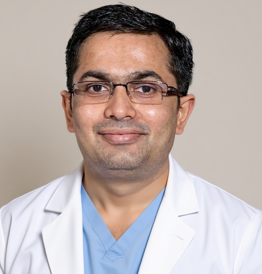
Dr Harsh Shah
MS, MCh (Surgical Gastroenterology), DrNB (Surgical Gastroenterology)
Intestine Cancer Surgeon
· Apollo Hospital, Bhat, Gandhinagar · Gota OPD, Ahmedabad
Qualified twice over in surgical gastroenterology, and operating in a part of India where a small bowel stricture is more often tuberculosis than cancer. That is not an academic distinction — it decides whether a patient keeps their intestine. Dr Harsh Shah takes tissue and waits for the answer in every case where the diagnosis is not already certain, including in the emergency theatre. Google rating 4.89 from 216 reviews at the Gota clinic, and 4.94 from 17 reviews at the Apollo, Bhat listing.
The small intestine is the long middle section of the digestive tract, between the stomach and the colon. Cancer here is rare, and it is not one disease. Four different tumours arise in the same organ — adenocarcinoma, neuroendocrine tumour, GIST and lymphoma — and each is treated completely differently. Naming which one it is comes before planning anything.
Adenocarcinoma is the tumour most people mean by small bowel cancer, and it is the one surgery is aimed at. Neuroendocrine tumours are slow-growing and, in many hospital series, actually the commonest malignancy found in this part of the bowel; they are removed surgically but behave quite unlike an adenocarcinoma afterwards. GIST arises from the wall rather than the lining and responds to a targeted tablet. Lymphoma is a blood cancer that happens to grow in the bowel, and it is treated by haemato-oncologists with chemotherapy — an operation for it is usually a mistake.
Tumours of the duodenum and ampulla, at the very top of the small intestine, are assessed separately — they involve the bile duct and pancreas and need a different operation.
In this country, a mass or a stricture in the small intestine is more often intestinal tuberculosis or lymphoma than it is cancer. Both can produce months of colicky pain, weight loss and recurrent obstruction, and both can look identical on a CT scan. Tuberculosis is cured by a full course of tablets. Lymphoma is treated with chemotherapy. Removing bowel for either of them is harm that a biopsy would have prevented.
It is why Dr Harsh Shah will often slow a plan down rather than speed it up. Tissue goes not only for ordinary histology but for AFB staining, GeneXpert and mycobacterial culture — the tuberculosis panel — because histology alone can miss it. Necrotic mesenteric nodes on the CT push the suspicion towards tuberculosis; bulky nodes, a dilated loop that is not actually obstructing, night sweats and a raised LDH push it towards lymphoma. Crohn disease, with its skip lesions and long history, is the third impostor.
The same rule applies in an emergency. When a patient arrives obstructed or perforated and an unexpected mass is found, tissue is taken at that operation and the full panel sent, because that tissue is only obtainable once. A resection done blind, on an assumption, cannot be undone.
The small intestine is the part of the gut that a gastroscopy and a colonoscopy both miss. If you have iron-deficiency anaemia or bleeding you cannot account for, and both of those scopes were normal, the next test is small bowel imaging — not a third repeat of the same two endoscopies.
That one rule shortens the delay that defines this disease. Small bowel cancer is typically diagnosed months after the symptoms start, because intermittent colicky pain is labelled functional and recurrent partial obstruction is treated and sent home. Very often the diagnosis is finally made on the operating table, during surgery for an obstruction nobody had explained.
| Step | Test | What it answers |
|---|---|---|
| 1 | CT or MR enterography | The primary test — is there a mass or a stricture, where is it, and are the mesenteric nodes necrotic? |
| 2 | Device-assisted enteroscopy | Reaches the lesion to take tissue and tattoo it, when it is within reach |
| 3 | Capsule endoscopy | For unexplained bleeding — only after obstruction has been excluded on imaging, because a capsule can lodge in a stricture |
| 4 | Staging CT of chest, abdomen and pelvis; CEA and CA 19-9 | Has it spread, and what is the baseline to follow afterwards |
| 5 | MMR or MSI testing on the tumour | Identifies Lynch syndrome, and identifies the patients for whom immunotherapy works |
One further step belongs in every case: a deliberate search for a condition that caused the cancer — Crohn disease, coeliac disease, Lynch syndrome, familial adenomatous polyposis or Peutz-Jeghers syndrome.
Four situations where an operation is the wrong answer: the diagnosis turns out to be tuberculosis, which needs a full course of anti-tuberculous tablets; it turns out to be lymphoma, which needs chemotherapy; it turns out to be Crohn disease, which is managed medically; or the cancer has already spread widely, where chemotherapy and relief of the blockage achieve more than a heroic operation would.
The fourth is worth saying plainly. Where disease is found scattered through the abdomen at operation, the right response is to biopsy it, relieve the obstruction with a bypass or a stoma, and stop. Attempting to clear everything there causes harm without buying time.
There is a fifth, quieter one. Where the diagnosis is uncertain and the patient is stable, waiting for the tissue answer is itself a decision, and the correct one. Dr Harsh Shah would rather explain a two-week delay than a resected intestine that turned out to be treatable with tablets.
For small bowel adenocarcinoma, removing the affected segment is the only treatment that cures. The segment comes out together with a wide wedge of the mesentery beside it, so that enough lymph nodes are removed for the pathologist to stage the disease accurately. A tumour sitting close to the caecum is dealt with by a right hemicolectomy instead, to get proper nodal clearance.
Two things are done at every operation regardless of the tumour. The whole small intestine is run through the surgeon’s hands from end to end, because a second tumour elsewhere is not rare — particularly in neuroendocrine tumours and in the inherited syndromes. And the length of bowel removed and the length left behind are recorded, because that number governs nutrition and vitamin replacement for the rest of the patient’s life. Where the tumour is stuck to something next to it, that structure is taken with it in one piece rather than peeled away. Peeling risks leaving cancer cells behind.
The approach — open or laparoscopic — is chosen for the tumour and the patient, not for the brochure. A small, localised tumour in a stable patient is often done laparoscopically and recovery is quicker for it; a bulky tumour, dense adhesions or an emergency perforation is done open, and that is a judgement, not a failure.
Preparation matters more here than in most operations, because these patients arrive malnourished after months of partial obstruction. Nutrition is built up first, anaemia corrected, sugars controlled, and a stoma site marked wherever bowel might be removed — even when a stoma is unlikely. Marking it costs nothing; needing one unmarked costs the patient a badly placed bag.
Chemotherapy after surgery is offered when the lymph nodes are involved, and discussed where the tumour had other high-risk features. For disease that has already spread, chemotherapy is the main treatment. And where the tumour is MMR-deficient or MSI-high, immunotherapy works remarkably well — which is why that test is done on everybody, not on a selected few.
Small bowel adenocarcinoma is biologically its own disease, and treating it as though it were colon cancer has already cost patients. A class of targeted drugs adopted straight from colorectal practice, the EGFR inhibitors, has not shown clear benefit here and is moving out of use. Taxane chemotherapy has moved in instead. The practical consequence for a patient is simple: ask whether the plan was written for small bowel cancer specifically, or borrowed from a commoner disease.
Because this cancer is uncommon, every case here goes to the tumour board — surgeon, medical oncologist, radiologist and pathologist together — and so does every case where tuberculosis or lymphoma has not been definitively ruled out.
★★★★★
“I am writing to express my sincere gratitude for the exceptional care provided by Dr. Harsh Shah and his team. Dr. Harsh Shah is an outstanding expert in intestinal surgery, executing complex procedures with remarkable calm and precision. Dealing with gastrointestinal diseases is incredibly stressful for both patients and their families. In my experience, what sets Dr. Harsh Shah apart is his incredible patience; he takes the time to listen to every question and provides clear, reassuring answers to every concern. As a grateful patient, I highly commend Dr. Harsh Shah and the entire team at Apollo Hospital. They did not just treat our physical condition—they truly cared for our mental and emotional well-being" — Dipen Shah, 5 stars, Dr Harsh Shah – Robotic GI Surgeon, Apollo, Bhat, Gandhinagar”
An unedited Google review from a patient treated for intestinal disease by Dr Harsh Shah, quoted word for word from the listings, with the original spelling and phrasing.
From the first consultation to full recovery there are five stages, and at each one you are told what happens next and when.
No single figure covers this. A patient whose tissue turns out to be tuberculosis is treated with tablets for months and never has an operation at all; a patient who needs a resection, and possibly chemotherapy afterwards, is on a different path entirely. What can be given is a written estimate for each stage before it happens, and a cashless pre-authorisation where the policy allows.
What moves the estimate: whether the admission is planned or an emergency, how much nutritional build-up is needed first, the approach used, whether a stoma is formed, the length of stay and the room category, and whether chemotherapy follows. An emergency admission costs more and carries more risk than the same operation planned — one more reason not to let a diagnosis drift.
The office prepares and submits the pre-authorisation before each admission. Bring the policy document and card, photo identity, the scans on disc, the biopsy and blood reports, and a list of your medicines with doses.
Not on its own. Intestinal tuberculosis, lymphoma and Crohn disease all produce the same picture, and all three are treated with medicines. A CT scan cannot reliably separate them from cancer — tissue can, provided the tuberculosis panel is sent alongside ordinary histology.
Because both standard scopes miss this part of the gut, and the symptoms are vague — intermittent colicky pain, episodes of partial blockage that settle, slow anaemia. Many patients are diagnosed only at an operation for obstruction.
Usually not. In a planned operation on a patient who has been nourished and prepared, the bowel is joined back together straight away. A stoma becomes likely when the operation is an emergency, when perforation has contaminated the abdomen, or when the patient is too malnourished for a join to heal safely.
The plan changes completely, and that is a good outcome. Anti-tuberculous treatment is started and the full course is completed — stopping early is what causes relapse and resistance. The case is notified and household contacts screened. This possibility is discussed before surgery, so a change in diagnosis afterwards is understood rather than alarming.
Most patients do — the small intestine is long, and removing a segment is usually well tolerated. Two things are watched after the ileum, the last part, has been removed: loose stools from bile-salt malabsorption, which a simple medicine treats, and loss of vitamin B12 absorption, which may mean lifelong B12 injections.
Sometimes, and it is worth knowing. Lynch syndrome, familial adenomatous polyposis, Peutz-Jeghers syndrome, coeliac disease and Crohn disease all raise the risk. Where a hereditary syndrome is found, genetic counselling and screening are offered to relatives — which can pick up disease in someone with no symptoms at all.
Shah’s Gastro, Cancer & Robotic Surgery Centre
Gota, Ahmedabad, Gujarat
Google rating 4.89 from 216 reviews
Dr Harsh Shah consults at two addresses in the Ahmedabad–Gandhinagar area: the clinic at Gota, Ahmedabad, and Apollo Hospital at Bhat, Gandhinagar.
Patients travel to Dr Harsh Shah from across Gujarat — Gandhinagar, Rajkot, Surat, Vadodara, Bhavnagar, Mehsana and Kutch — and from Rajasthan and Madhya Pradesh. For a suspected small bowel tumour, send the enterography on WhatsApp before travelling: if the images show something that needs tissue first, that can be arranged on the same visit rather than a second trip.
If you have been advised surgery for a small bowel mass, ask what the biopsy showed. If no tissue has been taken, that is the conversation to have before the operation is booked. Call +91-63555-64601 · WhatsApp Dr Harsh Shah's office
Swasth Parivar: twice a month, practical advice on recovery, diet and warning signs. No advertising. Stop any time.
Your number is used for these updates and for the clinic to reach you. It is never sold or passed on. To stop, reply STOP on WhatsApp.